Author: Nattha Wannissorn, PhD

  • Neurofeedback Part 2: Advantages, Side Effects, and Support Supplements

    Neurofeedback Part 2: Advantages, Side Effects, and Support Supplements

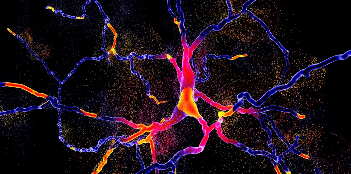
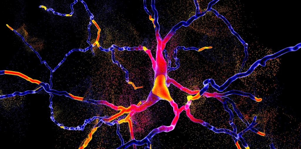
    Neurofeedback can be a powerful tool for brain optimization and changing your mental states. In the past article, part 1, we explained how it works and conditions that it’s been studied in. In this article, part 2, we’re discussing the advantages and side effects of neurofeedback, along with supplements that may help. 

    Advantages of Neurofeedback

    Non-Invasive and Drug-Free

    Neurofeedback is a non-invasive therapy that does not involve medication. This appeals to individuals seeking alternative or adjunctive treatments .

    Customized Treatment

    The therapy can be tailored to an individual’s specific needs by targeting certain brainwave patterns associated with their symptoms or goals .

    Potential Long-Term Benefits

    While research is ongoing, some users report lasting improvements after completing a course of neurofeedback sessions .

    Side Effects of Neurofeedback

    Neurofeedback is generally considered safe, but like any therapeutic intervention, it may have potential side effects for some individuals . It’s essential to note that these side effects are often mild and temporary. Here are some potential side effects of neurofeedback.

    1. Fatigue

    Some people may feel tired and sleepy after a neurofeedback session, especially during the initial stages of treatment.  Neurofeedback often involves concentration and mental effort as individuals work to modulate their brainwave patterns. The cognitive engagement required during the session can lead to mental fatigue .

    Furthermore, as the brain undergoes changes in response to neurofeedback, particularly in the modulation of specific brainwave frequencies, the adaptation process itself can be tiring. The brain is essentially learning to operate in a new or more optimized way, which may require additional energy

    2. Headache 

    Headaches can occur, particularly if the session involves a significant amount of concentration or if the individual is prone to headaches. Prolonged concentration can lead to tension headaches for some individuals .

    The visual and auditory stimuli used in neurofeedback sessions may trigger headaches in individuals who are sensitive to sensory stimuli or prone to migraines.

    People have varying levels of sensitivity to stimuli, and some individuals may be more prone to headaches in response to changes in visual or auditory stimuli used in neurofeedback.

    If neurofeedback involves the use of computer screens or other electronic devices, prolonged screen time can contribute to eye strain and, thus, headaches .

    3. Dizziness  

    Neurofeedback involves training the brain to modify its electrical activity patterns. As the brain adapts to new patterns during the session, some individuals may experience temporary dizziness as part of the adjustment process .

    Depending on the specific neurofeedback protocol, there may be deliberate efforts to alter certain brainwave frequencies. Rapid changes in these frequencies, particularly if significant, could contribute to sensations of dizziness .

    Neurofeedback sessions may involve visual or auditory stimuli. Individuals sensitive to such stimuli or with pre-existing sensory processing issues might be more prone to dizziness.

    Longer neurofeedback sessions or sessions with intense concentration may increase the likelihood of dizziness. This could be related to mental fatigue or strain .

    Some people are more susceptible to dizziness in response to changes in their sensory environment, and this sensitivity can influence their experience during neurofeedback.

    4. Emotional Changes 

    Neurofeedback can sometimes bring underlying emotions to the surface. As the brain undergoes changes in response to the training, individuals may experience shifts in emotional states. This emotional processing is a natural part of the therapeutic process.

    Certain neurofeedback protocols promote relaxation and stress reduction. As the brain learns to enter more relaxed states, individuals may experience changes in emotional well-being, including reduced anxiousness or increased calmness .

    5. Concentration Issues 

    Some people may find concentrating immediately after a neurofeedback session challenging, but this effect is usually short-lived .

    Neurofeedback involves training the brain to modify its electrical activity patterns. As the brain adapts to new patterns during the session, some individuals may experience temporary changes in cognitive processing, including concentration .

    Neurofeedback sessions, particularly those that are intensive or lengthy, may lead to mental fatigue. Fatigue can impact concentration.

    It’s crucial to communicate any side effects or concerns with the healthcare professional overseeing the neurofeedback sessions. They can adjust the protocol or provide additional support to minimize any adverse effects.

    Neurofeedback Support Supplements

    While neurofeedback is a non-invasive therapy, and its success doesn’t necessarily rely on specific supplements, some individuals may consider incorporating certain nutrients that are generally associated with brain health. 

    Intense neurofeedback sessions can also make you hungry or deplete more of certain nutrients. However, it’s important to consult with a healthcare professional before adding any supplements to your routine, as individual needs can vary, and some supplements may interact with medications or have contraindications.

    Here are some supplements that are commonly associated with brain health:

    1. Omega-3 Fatty Acids 

    Found in fish oil and certain nuts and seeds, omega-3 fatty acids, particularly DHA and EPA, are linked to cognitive function and overall brain health .

    • Omega-3 fatty acids, specifically DHA and EPA, are vital for cognitive function and overall brain health. They help form myelin sheaths and support BDNF levels.
    • DHA, highly concentrated in the brain, plays a crucial role in the structure of neural cell membranes.
    • These fatty acids contribute to neurotransmitter function, enhancing communication between nerve cells for improved cognitive processes like learning and memory.
    • Maintenance of structural integrity in neuronal membranes supports optimal synaptic function and neural signaling.
    • Neuroprotective properties of omega-3s contribute to shielding nerve cells from damage, potentially slowing neurodegenerative progression.
    • Improved blood flow in the brain and support brain development

    2. Vitamin B Complex 

    B vitamins, including B6, B9 (folate), and B12, play essential roles in brain function

    • B vitamins help regulate homocysteine levels. Elevated homocysteine is associated with an increased risk of cognitive decline and neurodegenerative disorders. Adequate B vitamin levels contribute to the breakdown of homocysteine.
    • B vitamins are involved in brain cell DNA synthesis and repair processes. This is essential for maintaining neurons’ integrity and proper functioning, supporting overall brain health.
    • B vitamins are crucial for methylation, a biochemical process involved in various functions, including neurotransmitter synthesis and DNA regulation. Proper methylation is essential for optimal cognitive function.
    • B vitamins play a key role in energy production from carbohydrates, fats, and proteins, providing the energy necessary for brain cells to function. Adequate energy supply is vital for cognitive processes such as concentration and mental alertness.
    • Some B vitamins, such as B6 and B12, have antioxidant properties, helping to reduce oxidative stress in the brain. This can protect neurons from damage caused by free radicals and support long-term cognitive health.

    3. Magnesium 

    It’s found in various foods, including nuts, seeds, and leafy green vegetables .

    • Magnesium modulates neurotransmitter release, influencing mood and cognitive functions.
    • It plays a role in NMDA receptor activation, crucial for synaptic plasticity and memory.
    • Magnesium exhibits inflammation-balancing properties, including in the brain.
    • It contributes to vasodilation, supporting optimal blood flow to the brain.
    • Magnesium helps regulate the stress response, promoting relaxation and mental well-being.
    • It stabilizes neuronal membranes, supporting overall neural health and function.

    4. Zinc 

    Zinc is an essential mineral involved in neurotransmitter function and overall brain health. It can be obtained from sources like meat, dairy, and legumes . Zinc plays a vital role in cognitive function through several mechanisms.

    • Zinc helps regulate neurotransmitters, including glutamate and dopamine, which are essential for cognitive processes such as learning and memory.
    • Zinc modulates synaptic plasticity, influencing the strength and efficacy of connections between nerve cells. This is crucial for adaptive changes in the brain associated with learning and memory.
    • Acting as an antioxidant, zinc helps protect neurons from oxidative stress and free radical damage, supporting overall brain health.
    • Zinc is involved in DNA synthesis and repair, essential for properly functioning and maintaining brain cells.
    • Zinc interacts with NMDA receptors, contributing to the receptors’ regulation in synaptic transmission and memory formation.
    • Zinc supports the generation of new neurons, a process known as neurogenesis, which is vital for cognitive function, especially in areas related to learning and memory.

    5. Vitamin D 

    Vitamin D has implications for brain health, and its deficiency has been associated with cognitive decline. Sunlight exposure and dietary sources like fatty fish and fortified foods are common ways to obtain vitamin D  .

    • Vitamin D influences the expression of genes related to neurotransmitter synthesis, supporting effective communication between nerve cells.
    • It plays a role in shielding neurons from inflammation and oxidative stress, contributing to brain health.
    • Vitamin D promotes the production of neurotrophic factors, supporting the survival and growth of neurons.
    • Deficiencies in vitamin D are linked to cognitive decline.

    6. Ginkgo Biloba 

    This herbal supplement is believed to have antioxidant properties and potential benefits for cognitive function

    • Ginkgo biloba acts as a vasodilator, enhancing blood flow to the brain for improved oxygen and nutrient delivery.
    • It exhibits antioxidant effects, protecting neurons from oxidative stress, a factor linked to cognitive decline.
    • Ginkgo biloba influences neurotransmitter activity, particularly acetylcholine levels associated with memory and learning.

    7. Curcumin (Turmeric) 

    Curcumin, the active compound in turmeric, has several cognitive benefits .

    • Curcumin, found in turmeric, possesses inflammation-balancing effects, potentially reducing neuroinflammation, a contributor to cognitive decline.
    • Its antioxidant properties may combat oxidative stress.
    • Curcumin may modulate pathways related to brain-derived neurotrophic factor (BDNF) production, crucial for neuronal growth and survival.

    8. L-Theanine 

    Found in tea leaves, L-theanine is an amino acid that may have calming effects and promote relaxation without sedation. It is often paired with caffeine for a balanced focus .

    • L-theanine induces a state of alert calmness by increasing alpha brainwave activity.
    • It positively affects neurotransmitters, particularly serotonin and dopamine, improving mood and focus.

    Conclusion:

    Neurofeedback can be a powerful tool to optimize your cognition and mental states, although the science is still in its infancy. It’s often advantageous to other modalities but not without side effects. In some cases, you can enhance neurofeedback benefits or reduce side effects significantly by providing your body with what it needs with food, sleep, and supplements. In other cases, supplements can boost the benefits of neurofeedback, especially for cognitive optimization.

    See also Part 1: What is neurofeedback? How does it work? What are the benefits?

    References:

    1. Allison, J., Kaliszewska, A., Uceda, S., Reiriz, M., & Arias, N. (2021). Targeting DNA Methylation in the Adult Brain through Diet. Nutrients, 13(11). https://doi.org/10.3390/nu13113979
    2. Calderón-Ospina, C. A., & Nava-Mesa, M. O. (2020). B Vitamins in the nervous system: Current knowledge of the biochemical modes of action and synergies of thiamine, pyridoxine, and cobalamin. CNS Neuroscience & Therapeutics, 26(1), 5–13.
    3. Calvello, R., Cianciulli, A., Nicolardi, G., De Nuccio, F., Giannotti, L., Salvatore, R., Porro, C., Trotta, T., Panaro, M. A., & Lofrumento, D. D. (2017). Vitamin D Treatment Attenuates Neuroinflammation and Dopaminergic Neurodegeneration in an Animal Model of Parkinson’s Disease, Shifting M1 to M2 Microglia Responses. Journal of Neuroimmune Pharmacology: The Official Journal of the Society on NeuroImmune Pharmacology, 12(2), 327–339.
    4. Chicago Mind Solutions. (2018, August 11). Are there any side effects to neurofeedback? Chicago Mind Solutions. https://chicagomindsolutions.com/are-there-any-side-effects-neurofeedback/
    5. Cuajungco, M. P., & Lees, G. J. (1997). Zinc metabolism in the brain: relevance to human neurodegenerative disorders. Neurobiology of Disease, 4(3-4), 137–169.
    6. Cuciureanu, M. D., & Vink, R. (2011). Magnesium and stress. University of Adelaide Press.
    7. Di Meo, F., Cuciniello, R., Margarucci, S., Bergamo, P., Petillo, O., Peluso, G., Filosa, S., & Crispi, S. (2020). Ginkgo biloba Prevents Oxidative Stress-Induced Apoptosis Blocking p53 Activation in Neuroblastoma Cells. Antioxidants (Basel, Switzerland), 9(4). https://doi.org/10.3390/antiox9040279
    8. DiNicolantonio, J. J., & O’Keefe, J. H. (2020). The Importance of Marine Omega-3s for Brain Development and the Prevention and Treatment of Behavior, Mood, and Other Brain Disorders. Nutrients, 12(8). https://doi.org/10.3390/nu12082333
    9. Eyles, D. W. (2021). Vitamin D: Brain and Behavior. JBMR plus, 5(1), e10419.
    10. Garcia, A., & Zanibbi, K. (2004). Homocysteine and cognitive function in elderly people. CMAJ: Canadian Medical Association Journal = Journal de l’Association Medicale Canadienne, 171(8), 897–904.
    11. Gómez-Pinilla, F. (2008). Brain foods: the effects of nutrients on brain function. Nature Reviews. Neuroscience, 9(7), 568–578.
    12. Kennedy, D. O. (2016). B Vitamins and the Brain: Mechanisms, Dose and Efficacy–A Review. Nutrients, 8(2), 68.
    13. Kirkland, A. E., Sarlo, G. L., & Holton, K. F. (2018). The Role of Magnesium in Neurological Disorders. Nutrients, 10(6). https://doi.org/10.3390/nu10060730
    14. Krall, R., Gale, J. R., Ross, M. M., Tzounopoulos, T., & Aizenman, E. (2022). Intracellular zinc signaling influences NMDA receptor function by enhancing the interaction of ZnT1 with GluN2A. Neuroscience Letters, 790, 136896.
    15. Kumar, V., Kumar, A., Singh, K., Avasthi, K., & Kim, J.-J. (2021). Neurobiology of zinc and its role in neurogenesis. European Journal of Nutrition, 60(1), 55–64.
    16. Layé, S., Nadjar, A., Joffre, C., & Bazinet, R. P. (2018). Anti-Inflammatory Effects of Omega-3 Fatty Acids in the Brain: Physiological Mechanisms and Relevance to Pharmacology. Pharmacological Reviews, 70(1), 12–38.
    17. Maier, J. A. M., Locatelli, L., Fedele, G., Cazzaniga, A., & Mazur, A. (2022). Magnesium and the Brain: A Focus on Neuroinflammation and Neurodegeneration. International Journal of Molecular Sciences, 24(1). https://doi.org/10.3390/ijms24010223
    18. Marzbani, H., Marateb, H. R., & Mansourian, M. (2016). Neurofeedback: A Comprehensive Review on System Design, Methodology and Clinical Applications. Basic and Clinical Neuroscience, 7(2), 143–158.
    19. Menon, V. P., & Sudheer, A. R. (2007). Antioxidant and anti-inflammatory properties of curcumin. Advances in Experimental Medicine and Biology, 595, 105–125.
    20. Nathan, P. J., Lu, K., Gray, M., & Oliver, C. (2006). The neuropharmacology of L-theanine(N-ethyl-L-glutamine): a possible neuroprotective and cognitive enhancing agent. Journal of Herbal Pharmacotherapy, 6(2), 21–30.
    21. Peng, Y., Ao, M., Dong, B., Jiang, Y., Yu, L., Chen, Z., Hu, C., & Xu, R. (2021). Anti-Inflammatory Effects of Curcumin in the Inflammatory Diseases: Status, Limitations and Countermeasures. Drug Design, Development and Therapy, 15, 4503–4525.
    22. Pickering, G., Mazur, A., Trousselard, M., Bienkowski, P., Yaltsewa, N., Amessou, M., Noah, L., & Pouteau, E. (2020). Magnesium Status and Stress: The Vicious Circle Concept Revisited. Nutrients, 12(12). https://doi.org/10.3390/nu12123672
    23. Prakash, A., Bharti, K., & Majeed, A. B. A. (2015). Zinc: indications in brain disorders. Fundamental & Clinical Pharmacology, 29(2), 131–149.
    24. Rogel, A., Guez, J., Getter, N., Keha, E., Cohen, T., Amor, T., & Todder, D. (2015). Transient Adverse Side Effects During Neurofeedback Training: A Randomized, Sham-Controlled, Double Blind Study. Applied Psychophysiology and Biofeedback, 40(3), 209–218.
    25. Sarraf, P., Parohan, M., Javanbakht, M. H., Ranji-Burachaloo, S., & Djalali, M. (2019). Short-term curcumin supplementation enhances serum brain-derived neurotrophic factor in adult men and women: a systematic review and dose-response meta-analysis of randomized controlled trials. Nutrition Research , 69, 1–8.
    26. Singh, S. K., Srivastav, S., Castellani, R. J., Plascencia-Villa, G., & Perry, G. (2019). Neuroprotective and Antioxidant Effect of Ginkgo biloba Extract Against AD and Other Neurological Disorders. Neurotherapeutics: The Journal of the American Society for Experimental NeuroTherapeutics, 16(3), 666–674.
    27. True Life Center. (2021, March 1). What are the neurofeedback side effects? True Life Center. https://www.truelifewellbeing.com/recovery-blog/what-are-the-neurofeedback-side-effects/
    28. Wang, L., Brennan, M., Li, S., Zhao, H., Lange, K. W., & Brennan, C. (2022). How does the tea L-theanine buffer stress and anxiety. Food Science and Human Wellness, 11(3), 467–475.
    29. Yamanaka, R., Shindo, Y., & Oka, K. (2019). Magnesium Is a Key Player in Neuronal Maturation and Neuropathology. International Journal of Molecular Sciences, 20(14). https://doi.org/10.3390/ijms20143439
    30. Yan, M., Song, Y., Wong, C. P., Hardin, K., & Ho, E. (2008). Zinc deficiency alters DNA damage response genes in normal human prostate epithelial cells. The Journal of Nutrition, 138(4), 667–673.
    31. Yoshitake, T., Yoshitake, S., & Kehr, J. (2010). The Ginkgo biloba extract EGb 761(R) and its main constituent flavonoids and ginkgolides increase extracellular dopamine levels in the rat prefrontal cortex. British Journal of Pharmacology, 159(3), 659–668.
  • Neurofeedback Part 1: How it works and benefits

    Neurofeedback Part 1: How it works and benefits

    If you’ve been serious about brain optimization, you may have heard of neurofeedback. In this article, part 1, we’ll cover everything you need to know about neurofeedback, from how it works to conditions that it’s been studied in. 

    In the next article, part 2, we’ll cover the advantages and side effects of neurofeedback, along with supplements that may make neurofeedback more effective.

    What Is Neurofeedback and How Does It Work?

    Neurofeedback is a concept that scientists have been studying for decades.
    Neurofeedback, also known as EEG (electroencephalogram) biofeedback, is:

    • A non-invasive therapeutic technique that helps train and regulate brain function
    • Based on the understanding that the brain’s electrical activity, or brainwaves as measured by EEG, reflects its functioning and can be modified to enhance mental health and performance
    • Allows you to control your brain waves and increase certain aspects of targeted brain activities through operant conditioning


    Our brains are made up of tiny cells called neurons that communicate with each other through electrical signals. When these neurons get active, they send out electrical pulses. Scientists can measure this activity by placing electrodes on the scalp, and the recording is called an EEG .

    The EEG shows different patterns of electrical activity, or brain waves, based on their amplitudes (power) and frequencies (how fast they oscillate). The frequencies are categorized into delta, theta, alpha, beta, and gamma waves, each linked to a specific function. 

    For instance, delta waves show up when a person is asleep, theta waves when they’re sleepy, alpha waves when they’re relaxed but awake, beta waves when they’re alert, and gamma waves when they’re trying to solve a problem .

    Neurofeedback is like a training session for your brain. You watch or listen to something, and your goal is to adjust your brain patterns based on what you see or hear. The focus of these training sessions is to learn to shift your thoughts and behaviors to produce a more desired pattern of brain wave activity. This allows the person to identify and prevent going back to the original unwanted brain activity patterns

    Here Is How Neurofeedback Works

    1. Assessment 

    A neurofeedback practitioner takes your physical, mental, and cognitive health history. They then initially assess your default brain pattern with EEG throughout your brain. 

    Based on this data, they may correlate the findings with known patterns with brain or mental health conditions, or specific physical health conditions. However, these EEG measurements in this context alone may not be diagnostic of any of these health conditions. 

    2. Goal Setting 

    Based on the assessment, your practitioner will help you set goals for the neurofeedback training. These goals are often related to optimizing certain brainwave patterns, such as improving cognitive function, emotional regulation, mood, empathy, discipline, or other targeted outcomes.

    3. Program Design

    Based on your goals and assessment, your neurofeedback practitioner will design your program. This involves:

    • Targeted brainwaves
    • Types of sensory inputs and feedback
    • Locations of electrodes
    • Frequency of training
    • Duration of sessions
    • Any other actions you need to take, such as avoiding coffee or certain foods, affirmations, exercise, and sleep requirements

    Many practitioners specialize in specific types of training or health conditions, so they may be more inclined to use the programs they’re familiar with. 

    3. Feedback Display

    During a neurofeedback session, you’ll receive visual or auditory feedback that reflects your current brainwave activity. This feedback is presented in real time, allowing the individual to observe and become aware of their brain’s patterns.

    4. Operant Conditioning 

    The individual is encouraged to consciously or unconsciously modify their brainwave activity to achieve the desired patterns. Positive feedback (e.g. visual changes on a screen or auditory signals) is provided when the brain produces the targeted patterns, reinforcing the desired changes.

    5. Repetition and Learning

    Through repeated sessions, the brain gradually learns to self-regulate and produce the desired patterns more consistently. This process is akin to training a muscle; the brain develops new, more adaptive neural pathways over time.

    6. Progress Monitoring

    Progress is continuously monitored either with EEG or other perceived changes, and adjustments to the neurofeedback protocol are made as needed. This ensures that the training remains tailored to the individual’s evolving brain activity.

    Neurofeedback Benefits

    While individual experiences may vary, here are some potential benefits associated with neurofeedback.

    Keep in mind that treating diseases with neurofeedback falls within the scope of practice for medical doctors and psychotherapists. You should see a neurofeedback-trained doctor if you have these conditions and are interested in treating them with neurofeedback. 

    1. Improved Focus and Attention

    Although research has produced mixed results, neurofeedback may help individuals enhance their ability to concentrate and maintain attention, making it particularly beneficial for conditions like ADHD .

    ADHD symptoms include inattention, distractibility, hyperactivity, and extreme dispassionateness. Neurofeedback therapy aims to normalize behavior without relying on medications that can have negative side effects .

    In ADHD treatment, the goal is to decrease brain activity in the theta band and increase activity in the beta band. Neurofeedback has shown long-term improvement in ADHD compared to traditional medications. Neurofeedback is effective in reducing hyperactivity, improving focus, academic performance, parental satisfaction, and indicators of sustained attention .

    In one study, 130 children with ADHD were randomly selected to receive either medication, neurofeedback and medication, or just neurofeedback treatment. Results collected included improvements in core ADHD symptoms, such as attention and hyperactivity, as reported by the parents .  

    According to this study, neurofeedback produced similar improvements as ADHD medications, highlighting the comparable efficacy of neurofeedback to a commonly prescribed medication. 

    The results suggest that neurofeedback can be a valuable alternative therapy for children and adolescents with ADHD, particularly the 20% who do not respond to medications. Additionally, the findings support the idea of reducing medication reliance when combining neurofeedback with other ADHD treatments .

    Another study on the efficacy of neurofeedback on adults with ADHD found mixed results. The study aimed to investigate the short and longer-term effects of two kinds of neurofeedback; SCP (slow cortical potential) and fNIRS (functional near-infrared spectroscopy) to a control group (EMG-BF). 

    All 84 participants in the study showed symptom improvements, but the fNIRS group, especially learners, demonstrated superior outcomes in ADHD symptoms and impulsivity ratings at the 6-month follow-up. 

    Neurofeedback involves learning to regulate brain activity. SCP focuses on slow electrical shifts in the brain, while fNIRS measures blood flow changes using light. The findings suggest that fNIRS neurofeedback, when learned effectively, can have specific and lasting benefits for adult ADHD .

    One double-blind, sham-controlled randomized controlled trial of fMRI neurofeedback for boys with ADHD found that neurofeedback made no improvements in ADHD. In fact, the sham group showed reduced irritable mood and improved motor inhibition post-treatment. The study suggests that fMRI neurofeedback may not be an effective treatment for ADHD

    Given the mixed effectiveness of neurofeedback on ADHD in the literature, it’s possible that individual responses to each type of neurofeedback may vary. This makes sense given that ADHD can vary widely from case to case.

    2. Mood Regulation and Enhancement 

    The inability to regulate your mood is linked to decreased metabolism in specific brain areas. Neurofeedback training targets increasing alpha and theta while inhibiting faster beta frequencies, significantly improving mood regulation .

    Training with neurofeedback has been linked to reduced stress levels, helping individuals achieve a calmer and more relaxed state . Alpha training, aiming to counteract the inhibitory effect of anxiety on alpha waves, is used to relieve anxiety symptoms .

    One study aimed to investigate the effect of mindfulness regulation through neurofeedback on 34 individuals; 17 of whom were diagnosed with anxiety disorder and the other 17 were healthy.

    The results showed a positive effect on both types of subjects. Specifically, there was an increase in the average power of gamma waves, which was interpreted as indicating relief from anxiety. The enhancement of wave power also suggested an improvement in the subjects’ mindfulness ability .

    The study suggests that neurofeedback mindfulness regulation can effectively influence the brain activity pattern of individuals with anxiety disorder, leading to relief from anxiety symptoms and improvements in mindfulness abilities .

    Another study assessed the efficacy of neurofeedback in individuals with chronic PTSD. 52 participants were randomized into neurofeedback and waitlist groups. Neurofeedback training occurred twice weekly for 12 weeks, targeting PTSD symptoms. Post-treatment, a significantly smaller proportion of the neurofeedback group (27.3%) met PTSD criteria compared to the waitlist group (68.2%). 

    There was a significant interaction between treatment condition and time, indicating the effectiveness of neurofeedback in reducing PTSD symptoms. The study concludes that neurofeedback warrants further investigation as a potential treatment for chronic PTSD, emphasizing its positive impact on symptom reduction and affect regulation capacities .

    3. Better Sleep

    Neurofeedback has demonstrated considerable potential in effectively addressing sleep-related challenges, particularly difficulty falling asleep. By actively contributing to the enhancement of sleep patterns, neurofeedback offers a promising avenue for individuals seeking not only faster sleep onset but also more restful and improved overall sleep quality .

    Although more research is still needed to evaluate the efficacy of neurofeedback in alleviating sleep disorders, several studies suggest neurofeedback improves sleep. However, such effects have also been attributed to possible placebo effects .

    One double-blind, placebo-controlled study tested the efficacy of neurofeedback on 25 patients with insomnia. One group received actual neurofeedback sessions, and the other group received sham treatments. Both neurofeedback and placebo feedback were found to be equally effective in improving subjective measures of sleep complaints


    The study did not find an advantage of neurofeedback over placebo feedback, and therefore, it could not be recommended as an alternative to cognitive behavioral therapy for sleep, which is considered the current (non-pharmacological) standard-of-care treatment

    A common beneficial side effect of neurofeedback is temporarily improved sleep on days of training. Possibly, this is because neurofeedback can be very taxing on the brain.

    4. Cognitive Enhancement and Language Skills

    Neurofeedback has emerged as a transformative influence in the realm of learning disability treatment, specifically for conditions such as dyslexia, characterized by challenges in reading and spelling, and dyscalculia, involving difficulties in mathematics. The therapeutic approach entails the augmentation of alpha wave activity to effectively target and ameliorate these learning disorders .

    Notably, individuals undergoing neurofeedback interventions have reported noteworthy enhancements in various cognitive domains. These include improvements in memory, heightened problem-solving skills, and an overall increase in mental clarity

    One study tested how neurofeedback treatment affects working memory (WM) in children with learning disorders. The study involved 18 children aged 8 to 11 years with learning disorders, they underwent either a neurofeedback treatment (NFB-group) or a placebo-sham treatment (sham-group) for 30 sessions. 

    Post-treatment, the NFB group demonstrated faster response times in the WM task. Additionally, there were changes in the WM-related EEG power spectrum, including decreased theta power and increased beta and gamma power at frontal and posterior sites, indicating improved efficiency of neural resource management, maintenance of memory representations, and enhanced subvocal memory rehearsal .

    Another study suggests that neurofeedback substantially improves spelling for dyslexic children. This study addressed reading and spelling deficits in dyslexic children through neurofeedback training based on neurophysiological differences. 

    Nineteen participants were randomized into an experimental group receiving neurofeedback and a control group, both receiving remedial teaching. The experimental group showed substantial spelling improvement

    However, a study comparing the efficacy of Cognitive Rehabilitation (CR) and neurofeedback found that neurofeedback is not as effective as CR in children with specific learning disorders. 53 elementary school students with Specific Learning Disorders were randomly assigned to neurofeedback, CR, or a control group. 

    After 20 sessions, the Continuous Performance Test (CPT) revealed a significant difference between groups, with the CR group outperforming neurofeedback. The study concludes that CR is more effective than neurofeedback in improving SA in students with Specific Learning Disorders .


    5. Addiction

    Neurofeedback may enhance the effectiveness of treatment for individuals struggling with substance abuse. While some studies note improvements in specific symptoms rather than overall quality of life or drug use, neurofeedback appears to address aspects of addiction beyond the reach of conventional treatments alone .

    One study treated ten adults addicted to cocaine with 12 neurofeedback sessions combined with motivational interviewing. The study did not have a specific control group. After the treatment, participants showed decreased reactions to drug-related images. 

    Clinical evaluations revealed reduced self-reported depression and stress, along with fewer instances of cocaine and marijuana use. The study suggests that neurofeedback has the potential to be a beneficial intervention for addiction .

    In another study, researchers randomly divided 93 participants with substance abuse disorders into two groups, one receiving 20 sessions of neurofeedback combined with the conventional treatment they were already receiving, and the other group only receiving conventional treatment. 

    Although the study did not find improvement in overall quality of life or reduction in drug use, it found that those receiving both neurofeedback as well as conventional treatment were less restless than those who only received conventional treatment. This study suggests that the use of neurofeedback can help alleviate symptoms that conventional treatment alone may not be able to target

    In a randomized, controlled trial researchers investigated the use of neurofeedback on 100 crystal methamphetamine-dependent (CMD) patients undergoing medical treatment. The treatment duration was two months, consisting of 30 sessions of neurofeedback for the experimental group, in addition to their usual medication. There was a control group that received only their usual medication. 

    The results showed that the experimental group had lower addiction severity, better psychological health, and improved quality of life compared to the control group. The differences between the two groups suggest that neurofeedback can enhance the effectiveness of treatment for CMD patients .

    6. Autism Spectrum Disorder (ASD)

    ASD is a neurodevelopmental condition characterized by enduring challenges that extend into adulthood. Children with ASD encounter obstacles in diverse domains, including social interactions, communication, and behavior, often exhibiting idiosyncratic behavioral patterns .

    Neurofeedback emerges as a valuable tool for supporting children with ASD. Studies show improvements in cognitive functions, social skills, and behavior after sessions. Tailored neurofeedback, guided by assessments, effectively addresses core ASD symptoms

    A study examining the efficacy of neurofeedback in treating ASD involved 42 children diagnosed with autism who underwent 40 neurofeedback sessions. After the therapy, the children showed improvements in their thinking, social skills, and attention. 

    The study suggests that neurofeedback could be a helpful treatment tool for reducing cognitive challenges in children with autism, indicating a positive potential for its effectiveness in enhancing their overall cognitive functions .

    In another study, researchers worked with two groups, one with ASD and another with typically developing (TD) children. The ASD group, consisting of 13 participants, received approximately 30 hours of neurofeedback sessions, while the TD group served as a control. The goal was to investigate changes in behavior and brain activity associated with social behaviors, particularly in the Mirror Neuron System (MNS). 

    The findings revealed that neurofeedback sessions had positive effects on the ASD group, showing improvements in behavior and increased engagement of the MNS. However, in the TD group, the results indicated a different response, suggesting that the impact of neurofeedback varied between children with ASD and typically developing children .

    These outcomes highlight the potential effectiveness of neurofeedback in improving social behavior in children with ASD, offering a promising avenue for intervention in neurodevelopmental conditions .

    Yet another study found that neurofeedback can be an effective treatment for ASD especially when guided by assessments. The controlled study involved 37 participants with ASD. The study found that those who received neurofeedback showed an 89% success rate in improving ASD symptoms, with significant improvements compared to the control group. 

    The treatment led to a 40% reduction in core ASD symptoms and a 76% decrease in hyperconnectivity, the presence of too many connections between different parts of the brain

    7. Epilepsy

    Neurofeedback may contribute to a reduction in the frequency and severity of seizures in individuals with epilepsy. It is proposed that by training individuals to regulate their brainwave patterns, specifically reducing abnormal electrical activity, neurofeedback can help minimize the occurrence of seizures

    Neurofeedback focuses on modulating specific brainwave frequencies. For epilepsy, the emphasis is on inhibiting abnormal patterns associated with seizures, such as slowing down theta waves or enhancing beta waves .

    A study explored whether two types of neurofeedback, SMR and slow cortical potentials (SCP), could help children and teens with controlled focal epilepsy. This study consisted of 44 participants with controlled focal epilepsy. There was a control group that received sham neurofeedback. 

    The participants were divided into three groups: one received sensorimotor rhythm (SMR) neurofeedback, another received SCP neurofeedback, and the third had sham neurofeedback. SMR neurofeedback trains individuals to regulate and control a specific brainwave that has an effect on attention and cognitive performance. 

    SCP neurofeedback works similarly, helping individuals regulate brainwave thought to influence neurological functions tied to attention and cognitive processes.

    The conclusions of the study suggest that SMR neurofeedback had positive effects on attention, both active and sham neurofeedback groups showed improvements in quality of life, and there was a trend for potential reduction in seizure frequency with SMR and SCP. 

    The study indicates that these neurofeedback approaches could positively affect cognitive performance, quality of life, and possibly seizures in children and adolescents with controlled focal epilepsy .

    8. Performance Enhancement 

    Neurofeedback training demonstrates promising benefits for athletic performance. It helps improve athletes’ skills, confidence, and overall performance in various sports .

    Researchers worked with seven healthy swimmers aged 18 to 25 over four months, having them undergo 20 neurofeedback training sessions. The results showed positive changes in the swimmers’ work curve during exercise, indicating improved mental performance. 

    Additionally, the study suggested that neurofeedback training impacted optimizing psychomotor activities. The changes observed were not huge, but they indicated trends toward better attention engagement during physical activities .

    Athletes who participate in sports that require high-level physical balance can benefit from neurofeedback. In one study, 18 judo athletes participated in a double-blind controlled experiment where some received neurofeedback training, and others did not. Those who received neurofeedback training significantly improved their dynamic balance


    In 2010, research involving Canadian athletes at the Vancouver Olympics revealed that employing neurofeedback (NFB) enhanced their ability to manage stress, leading to improved performance at the Olympics compared to the preceding year .

    Conclusion:

    Neurofeedback offers a non-invasive approach to enhancing mental health and performance. By training individuals to consciously control their brain waves, neurofeedback may help with a spectrum of conditions, from ADHD and mood regulation to sleep disorders and cognitive enhancement. 

    Remember that treating these conditions falls within the scope of practice for psychotherapists and medical doctors who employ neurofeedback as a tool in their toolboxes. Nowadays, there are many direct-to-consumer neurofeedback tools out there that promote well-being, calmness, and cognitive performance.

    The method involves real-time feedback, operant conditioning, and repetition, promoting neuroplasticity. While benefits include improved focus, mood regulation, and better sleep, individual experiences may vary

    Neurofeedback’s efficacy extends to diverse areas that involve changing brain functions and patterns. Moreover, it stands out for being non-invasive, customizable, and potentially yielding long-term benefits. As with any therapy, it’s important to address potential side effects and consider taking supplements for maximum support.
    Now that you’ve learned about what neurofeedback and what it helps with, check out part 2: neurofeedback advantages and side effects, and support supplements.

    References:

    1. Arns, M., & Kenemans, J. L. (2014). Neurofeedback in ADHD and insomnia: vigilance stabilization through sleep spindles and circadian networks. Neuroscience and Biobehavioral Reviews, 44, 183–194.

    2. Azizi, A., Mir Drikvand, F., & Sepahvani, M. A. (2020). Comparison of the Effect of Cognitive Rehabilitation and Neurofeedback on Sustained Attention Among Elementary School Students With Specific Learning Disorder: A Preliminary Randomized Controlled Clinical Trial. Basic and Clinical Neuroscience, 11(4), 465–472.

    3. Barth, B., Mayer-Carius, K., Strehl, U., Wyckoff, S. N., Haeussinger, F. B., Fallgatter, A. J., & Ehlis, A.-C. (2021). A randomized-controlled neurofeedback trial in adult attention-deficit/hyperactivity disorder. Scientific Reports, 11(1), 16873.

    4. Breteler, M. H. M., Arns, M., Peters, S., Giepmans, I., & Verhoeven, L. (2010). Improvements in spelling after QEEG-based neurofeedback in dyslexia: a randomized controlled treatment study. Applied Psychophysiology and Biofeedback, 35(1), 5–11.

    5. Chen, C., Xiao, X., Belkacem, A. N., Lu, L., Wang, X., Yi, W., Li, P., Wang, C., Sha, S., Zhao, X., & Ming, D. (2021). Efficacy Evaluation of Neurofeedback-Based Anxiety Relief. Frontiers in Neuroscience, 15, 758068.

    6. Coben, R., & Padolsky, I. (2007). Assessment-guided neurofeedback for autistic spectrum disorder. Journal of Neurotherapy, 11(1), 5–23.

    7. Duric, N. S., Assmus, J., Gundersen, D., & Elgen, I. B. (2012). Neurofeedback for the treatment of children and adolescents with ADHD: a randomized and controlled clinical trial using parental reports. BMC Psychiatry, 12, 107.

    8. Fotuhi, M. (2019, June 14). Neurofeedback: What it is and how it works | NeuroGrow. https://neurogrow.com/neurofeedback-complete-guide/

    9. Gabrielsen, K. B., Clausen, T., Haugland, S. H., Hollup, S. A., & Vederhus, J.-K. (2022). Infralow neurofeedback in the treatment of substance use disorders: a rando
      mized controlled trial. Journal of Psychiatry & Neuroscience: JPN, 47(3), E222–E229.
    10. Horrell, T., El-Baz, A., Baruth, J., Tasman, A., Sokhadze, G., Stewart, C., & Sokhadze, E. (2010). Neurofeedback Effects on Evoked and Induced EEG Gamma Band Reactivity to Drug-related Cues in Cocaine Addiction. Journal of Neurotherapy, 14(3), 195–216.

    11. Lambert-Beaudet, F., Journault, W.-G., Rudziavic Provençal, A., & Bastien, C. H. (2021). Neurofeedback for insomnia: Current state of research. World Journal of Psychiatry, 11(10), 897–914.

    12. Lam, S.-L., Criaud, M., Lukito, S., Westwood, S. J., Agbedjro, D., Kowalczyk, O. S., Curran, S., Barret, N., Abbott, C., Liang, H., Simonoff, E., Barker, G. J., Giampietro, V., & Rubia, K. (2022). Double-Blind, Sham-Controlled Randomized Trial Testing the Efficacy of fMRI Neurofeedback on Clinical and Cognitive Measures in Children With ADHD. The American Journal of Psychiatry, 179(12), 947–958.

    13. Luigjes, J., Breteler, R., Vanneste, S., & de Ridder, D. (2013). [Neuromodulation as an intervention for addiction: overview and future prospects]. Tijdschrift voor psychiatrie, 55(11), 841–852.

    14. Martínez-Briones, B. J., Bosch-Bayard, J., Biscay-Lirio, R. J., Silva-Pereyra, J., Albarrán-Cárdenas, L., & Fernández, T. (2021). Effects of Neurofeedback on the Working Memory of Children with Learning Disorders-An EEG Power-Spectrum Analysis. Brain Sciences, 11(7). https://doi.org/10.3390/brainsci11070957

    15. Marzbani, H., Marateb, H. R., & Mansourian, M. (2016). Neurofeedback: A Comprehensive Review on System Design, Methodology and Clinical Applications. Basic and Clinical Neuroscience, 7(2), 143–158.

    16. Maszczyk, A., Gołaś, A., Pietraszewski, P., Kowalczyk, M., Cięszczyk, P., Kochanowicz, A., Smółka, W., & Zając, A. (2018). Neurofeedback for the enhancement of dynamic balance of judokas. Biology of Sport / Institute of Sport, 35(1), 99–102.

    17. Mekkawy, L. (2021). Efficacy of neurofeedback as a treatment modality for children in the autistic spectrum. Bulletin of the National Salmon Resources Center , 45(1), 45.

    18. Mikicin, M., Mróz, A., Karczewska-Lindinger, M., Malinowska, K., Mastalerz, A., & Kowalczyk, M. (2020). Effect of the Neurofeedback-EEG Training During Physical Exercise on the Range of Mental Work Performance and Individual Physiological Parameters in Swimmers. Applied Psychophysiology and Biofeedback, 45(2), 49–55.

    19. Morales-Quezada, L., Martinez, D., El-Hagrassy, M. M., Kaptchuk, T. J., Sterman, M. B., & Yeh, G. Y. (2019). Neurofeedback impacts cognition and quality of life in pediatric focal epilepsy: An exploratory randomized double-blinded sham-controlled trial. Epilepsy & Behavior: E&B, 101(Pt A), 106570.

    20. Neurofeedback. (n.d.). Psychology Today. Retrieved January 26, 2024, from https://www.psychologytoday.com/us/therapy-types/neurofeedback

    21. Orndorff-Plunkett, F., Singh, F., Aragón, O. R., & Pineda, J. A. (2017). Assessing the Effectiveness of Neurofeedback Training in the Context of Clinical and Social Neuroscience. Brain Sciences, 7(8). https://doi.org/10.3390/brainsci7080095

    22. Pineda, J. A., Carrasco, K., Datko, M., Pillen, S., & Schalles, M. (2014). Neurofeedback training produces normalization in behavioural and electrophysiological measures of high-functioning autism. Philosophical Transactions of the Royal Society of London. Series B, Biological Sciences, 369(1644), 20130183.

    23. Rostami, R., & Dehghani-Arani, F. (2015). Neurofeedback Training as a New Method in Treatment of Crystal Methamphetamine Dependent Patients: A Preliminary Study. Applied Psychophysiology and Biofeedback, 40(3), 151–161.

    24. Rydzik, Ł., Wąsacz, W., Ambroży, T., Javdaneh, N., Brydak, K., & Kopańska, M. (2023). The Use of Neurofeedback in Sports Training: Systematic Review. Brain Sciences, 13(4). https://doi.org/10.3390/brainsci13040660

    25. Schabus, M., Griessenberger, H., Gnjezda, M.-T., Heib, D. P. J., Wislowska, M., & Hoedlmoser, K. (2017). Better than sham? A double-blind placebo-controlled neurofeedback study in primary insomnia. Brain: A Journal of Neurology, 140(4), 1041–1052.

    26. van der Kolk, B. A., Hodgdon, H., Gapen, M., Musicaro, R., Suvak, M. K., Hamlin, E., & Spinazzola, J. (2016). A Randomized Controlled Study of Neurofeedback for Chronic PTSD. PloS One, 11(12), e0166752.
    27. What happens during a neurofeedback session? (2019, July 18). Brainworks Neurofeedback | The UK’s Neurofeedback Leaders since 2007. London Clinic, Home Training and Industry Software Developers; Brainworks Neurotherapy. https://brainworksneurotherapy.com/about/neurofeedback-sessions/

  • What are Cognitive Functions and How Can You Boost Them?

    What are Cognitive Functions and How Can You Boost Them?

    Without them, you would struggle to:

    • Receive and process information
    • Store information
    • Remember information (short and long-term)

    Being able to do these things helps you manage the world around us. Your cognitive functions decline naturally as you age. You might already be experiencing this. For example, how sensitive we are to sounds begins to decline at age 30.

    However, you can do a lot to help support your cognitive function and stay sharp in the long run. In this article, we’re going to dig deep into the different cognitive functions and provide some tips on how to boost them. 

    In this article, Part 1, we’ll explain the various cognitive functions and how they work. In the next article, Part 2, we’ll cover ways to boost your cognitive functions.

    What Are The Different Cognitive Functions?

    Cognitive functions are a wide range of mental abilities. Let’s dive into each one and explain how they work. 

    Perception

    Perception is your brain’s interpretation of the world around you. This includes all of the information you take in via your five senses. Is it the same thing as reality? No. Interestingly enough, the activity in your brain is the same when you perceive something as when you hallucinate.

    Do you remember that dress that went viral a few years ago? People couldn’t decide whether it was white and gold or blue and black. That’s because how we perceive things differs from those around us. 

    So what’s happening neurologically to help us perceive and make sense of the world around us? Essentially, our brains make up a story about what we experience.

    Each of your senses has specialized nerve cells that turn the energy they receive into a neural signal and send it to the brain, making up a process called sensory transduction.

    Two neurotransmitters identified as responsible for your perception are dopamine and serotonin, both considered “feel good” chemicals. 

    In a study of 5 subjects, researchers explored the role of the dopamine and serotonin systems on perception and decision-making. Researchers recorded dopamine and serotonin levels while subjects undergoing deep brain stimulation played a computer game.

    Researchers observed that while subjects were perceiving dots on a screen, serotonin levels increased in the striatum, a part of your forebrain necessary for controlling voluntary movement when you feel uncertain. Right before subjects took action, dopamine levels increased while serotonin levels decreased.

    These results highlight the role of serotonin in slowing you down when you perceive information and the role of dopamine in speeding you up when uncertainty begins to fade. 

    What’s also interesting about perception is that while we do see things and make decisions, we don’t process this information in the same space as we do images. Perception occurs in the forebrain, while image processing occurs in the visual cortex at the back of the brain.

    Perception goes well beyond what you simply see. It’s how you process the information around you through your individual lens. 

    Memory

    Memory is probably the most well-known cognitive function. In its simplest form, it is the retention of information over time. It also allows you to recall information from the past and use it to understand the present. 

    When you think of memory, you’re probably thinking of long-term memory. It’s this type of memory that’s most worrisome when you notice you can’t remember that person’s name or recall certain words or phrases.

    There are two types of long-term memory:

    1. Explicit: events and facts
    2. Implicit: skills and habit – information that you gain and retrieve subconsciously

    You also have short-term memory, sometimes referred to as your working memory. It’s used to store information for a brief duration. For example, memorizing the access code in your email to input the information in an app or website. 

    So how do all of these memories become memories? Just like perception, it begins with the intake of sensory information. This information then travels into your prefrontal cortex where it stays for you to use as part of your short-term memory.

    For memories to become long-term, your hippocampus gets involved by retrieving information from your working memory. Then from there it changes your neural wiring to hold onto the information more permanently in the cerebral cortex. The hippocampus helps you connect your various sensory experiences and a particular event. It even works to layer on connections between new and past events. 

    Your brain has a particular region called the amygdala, an almond-shaped structure in the front of your temporal lobe behind your ears. The amygdala is responsible for your emotional memories. It is mainly activated when you’re presented with a threat or something that causes fear. The amygdala guarantees you remember precisely where you were and what you were doing on 9/11. 

    Also central to emotional memory is your cerebellum. It was previously thought only to regulate movement memory, but more recent research determined that the cerebellum is directly connected to the prefrontal cortex. The two parts of the brain work together to correct errors in movement based on memory, like correcting your basketball shot to improve your accuracy.

    Neurotransmitters that make memories happen include:

    • GABA (gamma-amino-butyric-acid): critical for memory consolidation, spatial memory, and memory retrieval
    • Acetylcholine: released in various areas of the brain when you experience something new or significant and when consolidating long-term memories
    • Glutamate: plays a critical role in memory formation by strengthening the connection between neurons
    • Dopamine: involved in the encoding and consolidation of new memories.
    • Noradrenaline: primarily active in the amygdala, noradrenaline interacts with cortisol (a “stress hormone”) to support emotional memory, processing, storage, and retrieval. 

    Learning

    Memory and learning are intricately linked. We learn through making memories. When you learn something new, just like memories your 85-plus billion neurons begin to fire and send electrical impulses to other neurons. These electrical signals allow you to read, write, think, and play sports. 

    These connections made in your brain occur because of neuroplasticity. It’s the idea that changes can occur in your brain and new connections (synapses) are made. These connections become more robust and faster through repetition. While you sleep, your brain prunes the extra or unused connections to become more efficient. 

    Learning and memory use the same parts of the brain and neurotransmitters. In the scientific world, learning is generally thought of as acquiring information that changes knowledge or behavior, while memory is the storage of information.

    Attention And Focus

    The modern world constantly bombards us with sensory information. Sounds and colors surround us. The internet is filled with competing news stories and social media reels. But our brain has limits. Our brains use focus and attention to filter important information and ignore the rest. 

    Driving your attention is either:

    • Goals: known as endogenous attention, where your brain is in charge of what you’re focusing on, or..
    • Stimulus: known as exogenous attention, where the external stimuli of your surrounding environment drive your attention

    Your brain allows you to switch back and forth between these two types of attention and to multitask by dividing your attention between the two.

    Your attention requirements change based on what you are trying to achieve. If you’re going for a walk down the street, you can let your mind wander and take in the sights and sounds around you. If you are trying to complete a work task, you’ll need to achieve mental focus or sustained attention.

    Your attention system in the brain is a connected network throughout almost every lobe in your brain. At the center are three different regions:

    1. Sensory regions: These include the primary auditory cortex, primary somatosensory cortex, and primary visual cortex and are responsible for processing incoming information. 
    2. Parietal regions: Located in the upper back portion of your skull behind your frontal lobe, this region controls where you should focus your attention.
    3. Prefrontal regions: Serves as your attention control center, particularly finding the balance between your endogenous and exogenous attention. 

    What you are focusing on determines what region(s) are most activated. 

    Also, these five key neurotransmitters ensure that your attention goes to where you need it most, allowing you to pay attention.

    • Acetylcholine: Works throughout the brain to enhance focus by controlling neural activity across the three attention regions of the brain. It increases your neural signal based on what area of the brain requires the most activity.
    • Dopamine: Helps focus your attention mainly when dealing with familiar information such as movement and emotional responses. Two different types of dopamine receptors work throughout the brain to either excite or inhibit cell activity.
    • Norepinephrine: Promotes arousal or wakefulness to improve your focus or increase reaction times.
    • Glutamate: Necessary for proper neuronal signaling to help maintain attention and focus.
    • GABA: Plays a special role in visual attention by focusing on relevant stimuli while blocking out irrelevant information. 

    Decision Making

    From what to wear to what to eat to what to watch, your day is filled with decisions. And I just mentioned some you make consciously. You make other subconscious decisions almost constantly throughout the day. While various factors go into decision-making, you’ll group them into two primary ways:

    • Perceptual decision-making: You gather information from the sensory information around you and use that to make decisions. For example, If you are walking down a sidewalk and another person is coming, you’ll pay attention to where you think they will go so that you can avoid that path and a possible collision.
    • Reinforcement-guided decisions: You base these decisions on more comprehensive knowledge and the value of a particular action. You decide what to do based on what you believe will give you the greatest reward. 

    The two most critical parts of the brain in the decision-making process are the prefrontal cortex and the hippocampus. They interact to help get you the information you need.  Researchers created an algorithm to break it down into four steps.

    • Step 1: You receive sensory information from a stimulus that excites neurons in the hippocampus.
    • Step 2: A secondary set of stimuli arrives in the hippocampus, producing information as a neural response.
    • Step 3: The information produced in step 2 is then sent to the prefrontal cortex, which determines whether you need more information to make a decision. 
    • Step 4: Finally, the prefrontal cortex makes the preferred decision, and you take action. 

    Different parts of the basal ganglia also play critical roles in decision-making. Here are a few highlights:

    • Nucleus Accumbens: the crucial area for reward processing. Reward drives decisions involved in this area of the brain.
    • Dorsal Striatum: engaged in making decisions about movement and taking action.
    • Subthalamic Nucleus: serves as a “brake” in decision-making to help prevent impulsivity.

    After making a decision, your amygdala comes into play. Based on the outcome of your decision, you experience a positive or negative emotional response. The amygdala stores this information and uses it when making a similar future decision.

    A study examining decision-making tracked the brain activity of participants. Researchers determined that your brain makes a decision up to ten seconds before you become aware of it. They were even able to accurately predict the decision a subject was going to make based on brain activity.

    Pulling this all together are neurotransmitters. Let’s explore which ones help you make decisions. 

    • Dopamine: Helps you make risk vs reward decisions.
    • Serotonin: Works together with dopamine to determine risk vs reward, with a focus on uncertainty.

    More recently, a study exploring decision-making focused on the relationship between serotonin and dopamine during the decision-making process. Using deep brain stimulation, researchers were able to track these neurotransmitters’ levels while subjects made decisions.

    Beyond the risk vs. reward role of dopamine and serotonin, researchers learned that both neurotransmitters help you choose to act. When subjects made decisions, serotonin levels decreased, and dopamine levels increased.

    • Acetylcholine: Tells you to feel uncertain about a decision when you don’t have enough information to predict the outcome.

    Inhibition

    Your inhibition helps you survive. It prevents you from taking impulsive action when you aren’t likely to benefit from it or if it’s not in line with your character. This extends to your thoughts and emotions as well. In some cases, you need inhibition to go off autopilot to actually do the right things, such as following instructions or driving on the other side of the road.

    Green, Purple, Orange, Yellow

    Caption: The Stroop test measures your inhibition skills by asking you to tell the color of the text. Most people will default to reading the text.

    Those who struggle with inhibition are more likely to engage in risky behavior such as alcohol and drug abuse and risky sex. And those with overactive inhibition are more likely to experience feelings of anxiety and difficulty in new social situations.

    A literature review explored the neuroscience of inhibition. Previously, researchers thought inhibition took place in one region of the brain– the right inferior frontal gyrus, part of your frontal lobe. However, they now find that inhibition includes a broad network of activity.

    Researchers determined that the inhibitory network extends to the cortical and subcortical regions of the brain. These regions include the outer layer of your two cerebral hemispheres and deep into the gray and white matter. This network of connection between the frontal lobe and other brain regions is active and interacting during inhibition control.

    The primary neurotransmitter involved in inhibition is GABA. GABA, known as a calming neurotransmitter, exerts the same type of influence on your thoughts and behaviors by reducing the excitability of your neurons.

    Dopamine is likely involved in overactive inhibition. In a literature review exploring the role of dopamine in inhibition, researchers found that the interactions of dopamine between the prefrontal cortex and midbrain regions like the amygdala and striatum can result in increased inhibition and anxious behavior.

    Spatial Orientation

    One of the most complex cognitive functions is spatial orientation. It requires you to take in spatial information in your environment and make sense of it in an organized manner so that you can successfully navigate the space around you. 

    This helps you get from one place to another, whether by driving over to a friend’s house or making your way to the bathroom in the dark at night. If we didn’t have these skills, we would essentially go around in circles and never make it to our destination. 

    The parts of the brain responsible for spatial orientation are your hippocampus, amygdala, and parahippocampus (located just outside the hippocampus). Within these regions are special cells with neurons specific to different types of information. They include:

    • Place cells: serve as a basis for cognitive maps, or maps that your brain creates to make sense of your surroundings, including specific landmarks. 
    • Head direction cells: help your brain orient the exact direction of your head independently of your surrounding environment
    • Grid cells: when these cells are firing, they can represent the entire spatial layout of your environment.
    • View cells: focus on individual objects or people in an environment

    A study examining the role of the hippocampus focused on taxi drivers as they navigated through a simulation of London streets. After undergoing MRI scans, researchers determined that compared to a control group, taxi drivers have larger hippocampuses. This demonstrates how critical of a role the hippocampus plays in spatial orientation.

    The most critical neurotransmitter for spatial orientation is acetylcholine. It influences how you experience what you see and integrate that information to make sense of it. It also allows you to focus your attention on specific visual stimuli, like zeroing in on a person or object of interest.

    Researchers were able to highlight the role of acetylcholine on spatial orientation by increasing acetylcholine in subjects using the drug donepezil. Compared to a control group, those with increased acetylcholine activity improved their ability to detect a target surrounded by distractions.

    The same study confirmed that enhancing dopamine and serotonin levels did not increase subjects’ ability to detect the target. This further confirms the critical role of acetylcholine in spatial orientation as opposed to other neurotransmitters.

    Language And Verbal Fluency

    Do you ever stumble over your words, trying to recall the name of something? Or feel like you’re lost in a conversation because of the vocabulary? This is a function of your language and verbal fluency. It includes your ability to:

    • Read
    • Write
    • Speak 
    • Communicate

    The language areas in the brain, located on the left side of the brain in the cerebral cortex, are aptly named after the neurologists who studied them, Paul Broca and Carl Wernicke. Broca’s area helps us recall words and speak, while Wernicke’s area decodes speech. 

    While the left side of the brain is primarily used in adults, a study focused on language development in children found something different. 73 subjects took verbal and fluency tests while tracking brain activity. Researchers found that how well children perform on verbal tests relies on the right hemisphere of the brain rather than the left. This is also true for adults.

    What we know about the neurotransmitters involved in language is a more recent discovery. Interestingly, scientists know less about the neurotransmitters involved in language than other cognitive functions because they cannot study language using animal models since animals don’t have language abilities. 

    The neurotransmitter extensively involved in language and verbal fluency is L-Glutamate. It plays a role in accessing, producing, and comprehending language. There are four different types of L- Glutamate receptors in the central nervous system, each of which plays its own unique role in overall fluency.

    The neurotransmitter GABA balances out L-Glutamate. GABA helps you slow down your speech and put pauses between your words and sentences. You can think of L-Glutamate as your language accelerator and GABA as the brakes.

    Now that we’ve covered your various cognitive functions, you may also be wondering if there are things you can do to boost them and ensure that you maintain high function. Of course, there are. In Part 2, we’ll cover 8 ways to boost your cognitive functions.

    References

    1. Murman DL. The impact of age on cognition. Semin Hear. 2015;36(3):111-121. doi:10.1055/s-0035-1555115
    2. Fish W. Perception, Hallucination, and Illusion. Oxford University Press; 2009.
    3. Butler AB. Cranial Nerves. In: Encyclopedia of the Human Brain. Elsevier; 2002:65-81.
    4. Bang D, Kishida KT, Lohrenz T, et al. Sub-second dopamine and serotonin signaling in human striatum during perceptual decision-making. Neuron. 2020;108(5):999-1010.e6. doi:10.1016/j.neuron.2020.09.015
    5. Ackerman S. From Perception to Attention. National Academies Press; 1992.
    6. Robertson LT. Memory and the brain. J Dent Educ. 2002;66(1):30-42. doi:10.1002/j.0022-0337.2002.66.1.tb03506.x
    7. Gao Z, Davis C, Thomas AM, et al. A cortico-cerebellar loop for motor planning. Nature. 2018;563(7729):113-116. doi:10.1038/s41586-018-0633-x
    8. Siegel SJ, Brose N, Janssen WG, et al. Regional, cellular, and ultrastructural distribution of N-methyl-D-aspartate receptor subunit 1 in monkey hippocampus. Proc Natl Acad Sci U S A. 1994;91(2):564-568. doi:10.1073/pnas.91.2.564
    9. Clos M, Bunzeck N, Sommer T. Dopamine is a double-edged sword: dopaminergic modulation enhances memory retrieval performance but impairs metacognition. Neuropsychopharmacology. 2019;44(3):555-563. doi:10.1038/s41386-018-0246-y
    10. van Stegeren AH. The role of the noradrenergic system in emotional memory. Acta Psychol (Amst). 2008;127(3):532-541. doi:10.1016/j.actpsy.2007.10.004
    11. Kania BF, Wrońska D, Zięba D. Introduction to neural plasticity mechanism. J Behav Brain Sci. 2017;07(02):41-49. doi:10.4236/jbbs.2017.72005
    12. Weiss JT, Donlea JM. Roles for sleep in neural and behavioral plasticity: Reviewing variation in the consequences of sleep loss. Front Behav Neurosci. 2021;15:777799. doi:10.3389/fnbeh.2021.777799
    13. Rothenfluh A, Troutwine BR, Ghezzi A, Atkinson NS. The genetics of alcohol responses of invertebrate model systems. In: Neurobiology of Alcohol Dependence. Elsevier; 2014:467-495.
    14. MacLean KA, Aichele SR, Bridwell DA, Mangun GR, Wojciulik E, Saron CD. Interactions between endogenous and exogenous attention during vigilance. Atten Percept Psychophys. 2009;71(5):1042-1058. doi:10.3758/APP.71.5.1042
    15. Petersen SE, Posner MI. The attention system of the human brain: 20 years after. Annu Rev Neurosci. 2012;35(1):73-89. doi:10.1146/annurev-neuro-062111-150525
    16. Dasilva M, Brandt C, Gotthardt S, Gieselmann MA, Distler C, Thiele A. Cell class-specific modulation of attentional signals by acetylcholine in macaque frontal eye field. Proc Natl Acad Sci U S A. 2019;116(40):20180-20189. doi:10.1073/pnas.1905413116
    17. Nieoullon A. Dopamine and the regulation of cognition and attention. Prog Neurobiol. 2002;67(1):53-83. doi:10.1016/s0301-0082(02)00011-4
    18. M. E, C. T, A. G, E. P. Dopamine and Glutamate Interactions in ADHD: Implications for the Future Neuropharmacology of ADHD. In: Banerjee S, ed. Attention Deficit Hyperactivity Disorder in Children and Adolescents. InTech; 2013.
    19. Leonte A, Colzato LS, Steenbergen L, Hommel B, Akyürek EG. Supplementation of gamma-aminobutyric acid (GABA) affects temporal, but not spatial visual attention. Brain Cogn. 2018;120:8-16. doi:10.1016/j.bandc.2017.11.004
    20. Khani A, Rainer G. Neural and neurochemical basis of reinforcement-guided decision making. J Neurophysiol. 2016;116(2):724-741. doi:10.1152/jn.01113.2015
    21. Saberi Moghadam S, Samsami Khodadad F, Khazaeinezhad V. An algorithmic model of decision making in the human brain. Basic Clin Neurosci. 2019;10(5):443-449. doi:10.32598/bcn.9.10.395
    22. Thibaut F. Basal ganglia play a crucial role in decision making. Dialogues Clin Neurosci. 2016;18(1):3. doi:10.31887/dcns.2016.18.1/fthibaut
    23. Gupta R, Koscik TR, Bechara A, Tranel D. The amygdala and decision-making. Neuropsychologia. 2011;49(4):760-766. doi:10.1016/j.neuropsychologia.2010.09.029
    24. Smith K. Brain makes decisions before you even know it. Nature. Published online 2008. doi:10.1038/news.2008.751
    25. Gunther KE, Pérez-Edgar K. Dopaminergic associations between behavioral inhibition, executive functioning, and anxiety in development. Dev Rev. 2021;60(100966):100966. doi:10.1016/j.dr.2021.100966
    26. Kang W, Hernández SP, Rahman MS, Voigt K, Malvaso A. Inhibitory control development: A network neuroscience perspective. Front Psychol. 2022;13:651547. doi:10.3389/fpsyg.2022.651547
    27. Bryson A, Hatch RJ, Zandt BJ, et al. GABA-mediated tonic inhibition differentially modulates gain in functional subtypes of cortical interneurons. Proc Natl Acad Sci U S A. 2020;117(6):3192-3202. doi:10.1073/pnas.1906369117
    28. Maguire EA, Woollett K, Spiers HJ. London taxi drivers and bus drivers: A structural MRI and neuropsychological analysis. Hippocampus. 2006;16(12):1091-1101. doi:10.1002/hipo.20233
    29. Gratton C, Yousef S, Aarts E, Wallace DL, D’Esposito M, Silver MA. Cholinergic, but not dopaminergic or noradrenergic, enhancement sharpens visual spatial perception in humans. J Neurosci. 2017;37(16):4405-4415. doi:10.1523/JNEUROSCI.2405-16.2017
    30. Gonzalez MR, Baaré WFC, Hagler DJ Jr, Archibald S, Vestergaard M, Madsen KS. Brain structure associations with phonemic and semantic fluency in typically-developing children. Dev Cogn Neurosci. 2021;50(100982):100982. doi:10.1016/j.dcn.2021.100982
    31. Li W, Kutas M, Gray JA, Hagerman RH, Olichney JM. The role of glutamate in language and language disorders – evidence from ERP and pharmacologic studies. Neurosci Biobehav Rev. 2020;119:217-241. doi:10.1016/j.neubiorev.2020.09.023
  • California Poppy Seed Extract

    California Poppy Seed Extract

    Recognized by its beautiful orange flowers, California Poppy (Eschscholzia californica) grows across North America. Native Americans utilized this plant in their daily meals and their medicine. In this article, we will cover how California Poppy improves sleep, helps you relax, and more.

    Alkaloids And Their Biosynthesis

    Benzylisoquinoline alkaloids are metabolites with a wide range of effects, including relaxation and discomfort relief. California Poppy plants produce these alkaloids.

    Sanguinarine is a major benzylisoquinoline alkaloid originating from California Poppy. This alkaloid inhibits bacterial growth, and is even an ingredient found in toothpaste . Other alkaloids extracted from Eschscholzia californica are californidine, escholtzine, and caryachine .

    WRKY transcription factors play a role in alkaloid biosynthesis across plant species. California Poppy possesses 50 WRKY genes, with different expression levels across plant parts .

    Benefits Of California Poppy Seed Extract

    Sleep

    California Poppy seed extract can improve your sleep. In a study, mice received between 100 and 200 mg/kg body weight of California Poppy extract. Doses above 100 mg/kg induced sleep in mice .

    The alkaloids act on GABA receptors. GABA is an inhibitory neurotransmitter which can promote sleep .

    A study included 22 individuals who struggled with sleep. They received a maximum of 4 pills every night for a month. The pills contained 80 mg of California Poppy extract and 32 mg of Valeriana officinalis extract.

    The results after one month were:

    • Sleep struggles decreased by 30%
    • Total duration of sleep increased by 30 minutes
    • The efficiency of sleep increased from 78% to almost 85%
    • Waking up during the night decreased by 25% .

    30 patients with similar sleep issues received 20 drops of either Eschscholzia californica or Passiflora incarnata (passionflower) extract at bedtime for 40 days. Individuals in the California Poppy group slept 59 minutes longer than the passionflower group .

    Calming Effects

    California Poppy seed extract promotes relaxation. In a mice study, California Poppy extract dosage of 25 mg/kg body weight induced calm behavior .

    Moreover, another study demonstrated similar results. Mice received 25 mg/kg body weight of California Poppy extract. The staircase test showed anti conflict effects in the California Poppy group. Additionally, the poppy seed group spent more time in the lit box during the light/dark choice test .

    Another study included 264 individuals. One group received a placebo, and the other group received tablets containing Crataegus oxyacantha and Eschscholtzia californica extracts for 3 months. Individuals experienced significant improvement in their mood. They also reported increased feelings of calmness .

    Reduces Discomfort

    California Poppy seeds impact discomfort relief. A mice study showed that supplementation with this plant extract reduced discomfort in mice. Animals performed less discomfort-reducing activities, such as twitching, jerking, and stretching .

    A rat study dealt with effects of California Poppy extract on discomfort perception and peripheral nervous system issues. The injection dose was between 100 and 300 mg/kg body weight.
    A single dose significantly reduced discomfort perception in rats. Thermal and other stimuli had no effects on rats, as California Poppy extract inhibited the aching sensations .

    Try Dream Optimizer, which contains California Poppy extract. Spray Dream Optimizer under your tongue and then swallow after 30 seconds. Feel the calming effects within minutes.

    Side Effects Of California Poppy Seed Extract 

    As California Poppy seed extract promotes relaxation and calmness, supplementation can cause slowed breathing and sleepiness.

    In the human study, the pill containing 80 mg of California Poppy extract (and one more ingredient) caused frequent urination in one participant .

    11.5% of participants who received Eschscholzia californica extract (plus another plant extract) had mild digestive issues in another human study .

    Conclusion:

    California Poppy seed extract is a safe and effective sleep and relaxation remedy. However, it’s not for everyone, as some people experience some side effects. When in doubt, consult a qualified naturopathic doctor before introducing a new supplement.

    References:

    1. Yamada Y, Hirakawa H, Hori K, et al. Comparative analysis using the draft genome sequence of California poppy (Eschscholzia californica) for exploring the candidate genes involved in benzylisoquinoline alkaloid biosynthesis. Biosci Biotechnol Biochem. 2021;85(4):851-859.
    2. Stefan Gafner, Birgit M. Dietz, Kerry L. McPhail, Ian M. Scott, Jan A. Glinski, Fiona E. Russell, Megan M. McCollom, Jason W. Budzinski, Brian C. Foster, Chantal Bergeron, Mee-Ra Rhyu, and Judy L. Bolton. Alkaloids from Eschscholzia californica and Their Capacity to Inhibit Binding of [3H]8-Hydroxy-2-(di-N-propylamino)tetralin to 5-HT1A Receptors in Vitro#. ACS Publications. Published online February 22, 2006. https://pubs.acs.org/doi/full/10.1021/np058114h
    3. Yamada Y, Nishida S, Shitan N, Sato F. Genome-Wide Profiling of WRKY Genes Involved in Benzylisoquinoline Alkaloid Biosynthesis in California Poppy (Eschscholzia californica). Front Plant Sci. 2021;12:699326.
    4. Rolland A, Fleurentin J, Lanhers MC, et al. Behavioural effects of the American traditional plant Eschscholzia californica: sedative and anxiolytic properties. Planta Med. 1991;57(3):212-216.
    5. Fedurco M, Gregorová J, Šebrlová K, et al. Modulatory Effects of Eschscholzia californica Alkaloids on Recombinant GABAA Receptors. Biochem Res Int. 2015;2015:617620.
    6. Abdellah SA, Berlin A, Blondeau C, et al. A combination of Eschscholtzia californica Cham. and Valeriana officinalis L. extracts for adjustment insomnia: A prospective observational study. Afr J Tradit Complement Altern Med. 2020;10(2):116-123.
    7. Californica MT, In PIMT. A study to compare the efficacy of Eschscholzia. Accessed April 5, 2023. https://www.homoeopathicjournal.com/articles/89/3-2-27-387.pdf
    8. Rolland A, Fleurentin J, Lanhers MC, Misslin R, Mortier F. Neurophysiological effects of an extract of Eschscholzia californica Cham. (Papaveraceae). Phytother Res. 2001;15(5):377-381.
    9. Hanus M, Lafon J, Mathieu M. Double-blind, randomised, placebo-controlled study to evaluate the efficacy and safety of a fixed combination containing two plant extracts (Crataegus oxyacantha and Eschscholtzia californica) and magnesium in mild-to-moderate anxiety disorders. Curr Med Res Opin. 2004;20(1):63-71.
    10. Vivoli E, Maidecchi A, Bilia AR, Galeotti N, Norcini M, Ghelardini C. Antihyperalgesic Effect of Eschscholzia Californica in Rat Models of Neuropathic Pain. Nat Prod Commun. 2008;3(12):1934578X0800301230.
  • What Are The 7 Major Neurotransmitters To Target With Nootropics?

    What Are The 7 Major Neurotransmitters To Target With Nootropics?

    It’s important to note that having a great night’s sleep is critical for the production of multiple neurochemicals.

    There are 7 key players in your brain’s chemical makeup.

    neurotransmitters

    1. Dopamine: The Molecule Of Drive And Motivation

    If you’ve been feeling overwhelmed and burnt out, with zero energy to get through your day, there could be another mischievous, lurking reason as to why that is. 

    It all has to do with your neurochemicals. Most notably, dopamine. When your levels of dopamine are rocking and rolling, life is pretty great. This is because dopamine is responsible for your body’s “reward” system that helps you focus. 

    So when you’ve got great levels of dopamine, you feel AWESOME as you work step by step towards your goals. It will reinforce behavior and make you feel good, like that giddy feeling you get when you’re looking forward to something exciting. 

    This in turn helps you get into GOOD habits, and avoid the low energy or “burn out” feeling. For example, say you know you’ve got to get in a morning run. When you’ve got good dopamine levels, you’ll think about something FUN in that run that will get you moving and grooving and ready to crush your routine. Dopamine even reinforces memories. Having good levels of dopamine is critical to success. 

    There are some natural ways to boost your dopamine:

    Sunlight: direct sunlight in the eyes boosts dopamine and serotonin.  Another reason to lock in that morning sunlight ritual.

    Cold therapy:  exposure to cold has been shown to boost dopamine by 250%.  That’s an incredible gain.

    Nootropics: nootropics are brain enhancing supplements that can directly level up your neurochemicals including dopamine.

    We created a personalized nootropic company that produces the most powerful nootropic stacks on the market.  We have several formulas that increase dopamine including: Powerful Solution, Apex, Dopa Drops and Zoned In.

    2. Acetylcholine: The Molecule Of Focus

    On a day-to-day basis, you have to rely on your BRAIN to maximize on all of the necessary steps that will get you to your end goal. This is true whether your goal is to triple your revenue growth, overcome your strongest competitor, etc.

    That’s why investing in your brain is one of the most critical things you can do for your career long term. With all of this in mind, it’s important to think about 3 questions:

    1. How long does it usually take you to think up new project ideas?
    2. How long does it take you to recall information?
    3. How long does it take you to learn something new?

    Imagine for a moment what your day would look like if you were able to INCREASE your ability to do all of those things at lightning speeds:

    • You would be able to come up with creative new project proposals faster, see results faster, and grow your business faster.
    • You would be able to boost your memory and attention span to accomplish double the amount you would have been able to before in a day’s work. 
    • And you would be able to learn more at faster speeds to reach your goals and rise above the competition.
    neurons

    The key to making this a reality is acetylcholine.  Your thinking, memory, focus, and attention all rely on acetylcholine.

    Eggs are the best source of choline which your body converts into acetylcholine.  

    Nootopia has several formulas that boost your acetylcholine on demand including Apex, Mental Reboot AM and Nectar X. 

    3. Oxytocin: The Bonding Molecule

    Oxytocin is one of the most important molecules for great health, especially for women.  Men thrive on testosterone and women thrive with oxytocin.

    The best part of oxytocin is IT’S FREE.  You can get a significant boost in oxytocin by simply hugging someone for 22+ seconds. 

    Kids and pets are powerful oxytocin generators.  Just playing with them will give you a potent boost in oxytocin which will have a very positive impact on your health and mental perspective.

    4. GABA: The Molecule Of Chill

    Do you find yourself getting easily irritated by daily frustrations like: 

    • Traffic
    • Having nowhere to park
    • Your coffee getting cold
    • Or your phone battery dying? 

    Do you feel like your brain is going a mile a minute with jittery nervousness, and you’re unable to get calm for big meetings or social interactions? 

    Would you like to tap into a state of mind where you feel groovy, ready to rock and roll, and able to maintain a positive attitude through anything your day throws at you? 

    If you answered YES… Odds are you are LOW or DEFICIENT in the neurochemical GABA. GABA is the anti-stress “molecule of chill.” It’s a critical neurochemical not only for keeping stress levels low and “chillaxed” levels high, but it also plays a major role in keeping all of your other neurochemicals BALANCED.

    sinapsis

    GABA is flexible. You can control your dopamine, serotonin, and acetylcholine levels with GABA to make them each act more effectively, or reduce it to avoid jitters.

    In other words, GABA is key for connectedness. When your GABA levels are optimal, everything works in harmony to bring you groovy vibes.

    The great news is that if your levels are currently low… you can get GABA flowing in just 2 minutes. 

    Nootopia has 2 powerful GABA boosting formulas: Zamner Juice and GABAlicious.

    5. Serotonin: The Molecule Of Stability

    If you’re low in serotonin, a couple things start to happen…

    1. You struggle to get into a good mood, always feeling blue
    2. You begin to lack the drive to do things you know you ought to be getting done
    3. Your brain becomes foggy 24/7
    4. And you no longer take pleasure in things you used to enjoy doing

    Low levels of serotonin are linked to depression, anxiety, and poor sleep. All of which keeps these 4 main negative impacts going in a vicious cycle. 

    That doesn’t sound like a very good way to enjoy life to the absolute fullest. So if you’ve started to notice some of these same traits in your own daily life…. 

    Getting sunlight is a powerful free way to boost your serotonin.

    Carbohydrates are another powerful way to increase serotonin.  This is one of the reasons why sugar and carbs can be additive.  

    Nootopia blends that can boost your serotonin include Upbeat, Zamner Juice and Nectar X.

    6. Noradrenaline: The Molecule Of Initiation

    Do you ever realize partway through a conversation that you’re not really “there” with that person? You feel a bit disconnected, or not very present? 

    Have you ever found yourself in the midst of a task when you realize you’re not very focused on what you’re actually doing? 

    Would you like to increase your wakefulness to boost your presence, reduce distractions, and pump out hard work effortlessly? 

    If you answered YES to any of these questions, then there’s a key neurochemical at play: Noradrenaline.

    Not to be confused with adrenaline, noradrenaline is responsible for arousal and wakefulness. We’re not talking about sexual arousal, but rather ALERTNESS and presence. 

    When your levels of noradrenaline are LOW, you feel:

    • A lack of energy
    • Inability to concentrate
    • Trouble focusing
    • And disconnected

    All of this adds up to an unproductive day with your task list piling up, and you’ll have trouble communicating and connecting with others and difficulty keeping your projects and deadlines straight.

    Not a great way to get to the next level of your career or increase your business growth.

    The GOOD news is your noradrenaline levels don’t have to stay that way. You CAN boost your noradrenaline to optimal levels.

    It’s a tricky neurochemical however, because TOO much and you’ll feel wired. You’ll burn out fast. You want to find that zone of magic where you’re in the sweet spot without overkill.That’s when you unlock a gentle extension of focused attention that lights up your brain, with the ability to control your focus throughout the day. 

    The best two Nootopia formulas for noradrenaline are Nectar X and Zoned In.

    neurons

    7. Adrenaline: The Molecule Of Aggression

    When you hear the word “adrenaline” what do you think of? Maybe you think of:

    • Stress
    • Fear 
    • Your heart racing
    • Feeling your heartbeat throbbing in your temples
    • Or rapid breathing

    Yes – Adrenaline is an important neurochemical for keeping us safe in dangerous moments where we need heightened senses or a flood of energy.  But many people don’t know how adrenaline can be used for SUCCESS.  The optimal levels of adrenaline can transfer your performance for athletic endeavors, workouts and other situations that require intensity and aggression.

    It’s possible to access adrenaline without tipping over into fear or the physical signs of stress. When you are in control of your adrenaline levels, some pretty incredible things happen:

    • You’re able to push through barriers
    • You rise to the next level of focus to push your projects forward
    • You surge with intensity and drive, key for any physical task…
    • And your mental drive gets combined with your physical capacity to DO IT

    Listening to aggressive music is one of the best free ways to activate adrenaline.  What type of music is best for adrenaline differs.  For some it’s rock and metal, others its EDM and gangster rap. 

    Many people including fighters, soldiers and athletes are able to generate adrenaline on demand.  They’ve learned how to activate it by switching into an aggressive, high-performance state.

    Nootopia has created effective stacks that will boost your adrenaline including Power Solution and Zoned In.

    Optimal adrenaline levels = More gets DONE. Now, if you’ve been struggling lately to get things done, there’s a 10 minute solution for that. 

  • Celastrus Paniculatus

    Celastrus Paniculatus

    Due to the alleged cognitive effects of Celastrus paniculatus, sometimes people call it the “elixir of intelligence” or the “elixir of life” in traditional Ayurvedic medicine. Evidence suggests that this plant, especially its oil, improves mental performance and memory, affects neurotransmitters, and helps with pain and inflammation. 

    Disclaimer: Most Celastrus studies so far are animal studies. However, we’ve found it an effective nootropic herb, especially with our proprietary extraction technologies.

    What Is Celastrus Paniculatus?

    Celastrus paniculatus is a tall, woody climber (sometimes known as a climbing shrub) of the Celastraceae family with yellow, corky bark . It grows throughout India and may reach heights of about 2000 meters. The plants have a variety of medicinal properties, including usage for:

    • Cognitive impairment
    • Sleep
    • Joint discomfort
    • And post-meal stomach discomfort

    The most often utilized plant portion is oil from the seeds (known as Jyothismati oil, JO), which contains different levels of the alkaloids celastrine and paniculatin. Jyothismati has a bitter flavor and a scorching potency. However, the seeds and seed oil are highly therapeutic. The seed oil is beneficial for massage, particularly in vata disorders such as back discomfort, facial muscle weakness, and joint issues .

    Note: We’re sharing this list of conditions based on Ayurvedic literature for educational purposes only. Our products contain specific extracts of Celastrus that are not intended to treat any diseases.

    What Is SuperCelastrusTM?

    SuperCelastrusTM are our set of proprietary extracts of Celastrus peniculatus, using a combination of:

    • Sonic and ultrasonic extraction
    • Lipid suspension and micronization technologies
    • Combining various parts of the seed and the plant at various stages of processing

    Our extraction technologies allow us to create SuperCelastrus blends that target dopamine, acetylcholine, serotonin, and GABA. 

    Health And Brain Benefits Of Celastrus Paniculatus

    Celastrus Paniculatus

    Celastrus’ Effects on Neurotransmitters 

    Clinical studies indicate that Celastrus paniculatus has two main effects on neurotransmitters: 

    1. Balancing their levels 
    2. Antioxidant and neuroprotective effect

    Celastrus paniculatus increases cognitive performance by acting on the acetylcholine level in the brain. It primarily balances neurotransmitters such as serotonin, epinephrine, and dopamine to optimize memory and learning in the brain. 

    Celastrus paniculatus also enhances mental wellbeing and promotes a healthy stress response due to its interaction with dopamine, serotonin and GABA receptors. It increases the levels of serotonin, dopamine and noradrenaline by blocking the activity of MAO-A. MAO-A is the enzyme that breaks these neurotransmitters down.

    In rats, Celastrus may lower both levels and turnover of noradrenaline, dopamine, and serotonin levels in the brain. According to the researchers, these effects may aid learning and memory enhancement .

    Increases Acetylcholine Levels

    C. paniculatus seed extract inhibits acetylcholinesterase, the enzyme that breaks down acetylcholine. Blocking this enzyme boosts acetylcholine levels in the brain, improving many aspects of cognitive function .

    Taking Celastrus oil for prolonged periods of time can lead to increased cognitive performance and improvements in memory retention. This is due to the fact that Celastrus paniculatus increases levels of acetylcholine in the brain.

    Improves Mental Performance And Memory During Stressful Situations

    Chronic stress can accelerate aging and neurodegeneration . Stress can also impair some aspects of brain function .

    Long-term stress can interfere with working memory, learning, and relaxation responses. According to some experts, Celastrus may help protect the brain from the detrimental effects of stress. Stressed rats that were administered Celastrus oil performed better on learning and memory tests . Another rat study discovered that Celastrus seed oil might boost mental function and memory

    Improves Myelination

    Celastrus paniculatus oil increases the concentration of proteins and phospholipids in the brain leading to the belief that it possibly increases myelination around neurons.

    Protects Neurons From Glutamate Toxicity

    Glutamate is a chemical that can cause neuronal cell death, Celastrus paniculatus protects the brain from glutamate induced toxicity by blocking NMDA receptors.  

    Counteracts And Protects Neurons From Oxidative Stress

    Furthermore, the antioxidants in Celastrus paniculatus reduce brain levels of the oxidative stress marker malondialdehyde. Some of its components scavenge hydrogen peroxide and superoxides inside the cells. Aside from counteracting the harmful effects of oxidative stress, it also simultaneously increases glutathione and catalase levels significantly .

    Inflammation-Balancing

    Studies on Celastrus paniculatus indicate possible anti-inflammatory effects. In addition, there is some indication that Celastrus extract components (sesquiterpene esters) may inhibit inflammatory chemicals such as NF-kB, nitric oxide, IL-6, and TNF-a .

    In mice, Celastrus extract had inflammation-balancing effects. In addition, Celastrus seed extract may also ease swelling and discomfort in animal studies .

    We need additional clinical studies to confirm this benefit of Celastrus.

    Dosage 

    Traditional doses of Celastrus paniculatus begin with 10 seeds consumed all at once. If no adverse effects occur, raise the amount to 15, then 20, and eventually 30 seeds.

    Due to a lack of clinical study, a safe and effective dosage of Celastrus is unknown. However, Celastrus supplements may contain 400-500 mg of Celastrus paniculatus powder with standard doses of 1 to 2 capsules per day.

    Much more study is required to find the best dosage of Celastrus paniculatus.

    Side Effects 

    Due to a lack of clinical studies, no side effects have been determined for the usage of Celastrus paniculatus.

    Celastrus has non-existent toxicity in our lab, as we’ve found its LD-50 to be very high. It also doesn’t cause tolerance over time, so chronic use wouldn’t be a concern.

    Celastrus paniculatus FAQ

    Is Celastrus Toxic?

    There is a lack of clinical studies investigating the health effects and safety of Celastrus paniculatus. However, animal studies have touched upon this topic.

    1. Sperm cell depletion and arrest of spermatogenesis was seen when rats received 0.2 ml of the extract in 0.2 ml of peanut oil every 48 hours for 30 days. Furthermore, liver lesions and focal necrosis occurred during the treatment but healed when celastrus supplementation was discontinued.
    2. Female albino mice received 300, 2000, or 5000 mg/kg of celastrus seed oil for 42 days. There were no signs of side effects (diarrhea, skin changes, tremors, coma, etc.), toxicity, or lethality caused by the mentioned doses.
    3. In our lab, SuperCelastrus, our proprietary extract of celastrus, has shown no toxicity due to very high LD-50. 

    Thus, Celastrus paniculatus seems safe to use. However, dosages in animal studies tend to be very high compared to doses humans take, and some health effects don’t carry over. Therefore, clinical evidence in humans is necessary. 

    What Are the Effects of Celastrus paniculatus Seed Oil?

    Celastrus seed oil shows various nootropic activities in mice and rats. 

    1. It inhibits acetylcholinesterase to increase acetylcholine and increase acetylcholine between neurons, thereby boosting memory performance.
    2. Celastrus compounds associate with central muscarinic receptors to enhance spatial memory in rats. 
    3. The seed oil also interacts with monoamine oxidase A to decrease its activity in mice, and induce mood-boosting effects. 
    4. The seed oil supports healthy stress response in mobility-restricted rats.
    5. While the chloroform extract showed strong antioxidant properties, the aqueous extract had highest inflammation-balancing activity.  
    6. The seed oil supports gastrointestinal health and protects the stomach lining in rats.
    7. Methanolic seed extract also supports healthy cholesterol levels.

    Keep in mind that while rodent studies led to many scientific discoveries, you are not a rodent. So, we need more clinical studies to confirm these benefits in humans.

    What Is the Common Name for Celastrus paniculatus?

    Here are some of the common names for Celastrus paniculatus:

    • “Black oil plant” because the seed oil is a very dark color
    • “Intellect tree” 
    • “Climbing staff plant” due to its climbing abilities which result in the plant reaching up to 10-18 m (32-59 feet) tall and having 25 cm (9.8 inches) wide stems
    • “The tree of life” in Indian culture, where it’s called “malkankani”. 
    • “Jyotishmati” is another name for celastrus in the Sanskrit language (coming from intellect – medha, and memory – smruti), which is often used to refer to the Celastrus paniculatus seed oil.

    What’s in the Celastrus paniculatus Leaves?

    Celastrus leaves are alternate, broad, and oval/elliptical, and have dentate margins. Compounds found in the leaf extract include steroids, terpenoids, alkaloids, saponins, carbohydrates, and phenolic compounds. Celastrus paniculatus leaves obtained from the forest areas of India also contain fixed oils (in all extract types), flavonoids (in the water extract), and triterpenoids (in water and ethanolic extract). An in vitro study of leaf ethanolic extract reported a total phenolic content of 125.6 mg/gm equivalent to gallic acid in 1 mg/ml of the leaf extract. 

    Furthermore, the methanolic extract of celastrus leaves inhibited the growth of Phytophthora capsici fungi, with inhibition being 100%. Celapanin, a sesquiterpene isolated from the ethanolic extract of celastrus, significantly inhibited the growth of S. aureus, and moderately of K. pneumoniae and P. aeruginosa.

    Why Is Celastrus paniculatus Called the Intellect Plant?

    You might wonder how celastrus got one of its common names – “the intellect plant”. “Jyotishmati” is another name for celastrus in the Sanskrit language. It comes from the words medha, which means intellect, and smruti, which means memory. This is because Celastrus paniculatus was used to improve intellect in traditional Ayurveda medicine. This plant was mentioned in the writings of Charaka, Vagbhata, and Sushruta as a mental health and intellect-boosting agent, with a special focus on the celastrus oil. The ancient prescriptions included celastrus oil mixed with cow’s ghee. It is because of the ancient Indian use of this plant that it became known as “the intellect plant’’.

    Bottom Line

    Celastrus paniculatus has various medical and therapeutic benefits, such as improving mental performance and memory, exerting certain effects on neurotransmitters, and potentially inflammation-balancing. Although most of these findings come from animal studies, Celastrus is a very popular herb in Ayurvedic medicine with a long history of safe use.

    References

    1. Lekha G, Mohan K, Samy IA. Effect of Celastrus paniculatus seed oil (Jyothismati oil) on acute and chronic immobilization stress induced in swiss albino mice. Pharmacognosy Res. 2010;2(3):169-174. doi:10.4103/0974-8490.65512
    2. Godkar PB, Gordon RK, Ravindran A, Doctor BP. Celastrus paniculatus seed water soluble extracts protect against glutamate toxicity in neuronal cultures from rat forebrain. J Ethnopharmacol. 2004;93(2-3):213-219. doi:10.1016/j.jep.2004.03.051
    3. Nalini K, Karanth KS, Rao A, Aroor AR. Effects of Celastrus paniculatus on passive avoidance performance and biogenic amine turnover in albino rats. J Ethnopharmacol. 1995;47(2):101-108. doi:10.1016/0378-8741(95)01264-e
    4. Bhanumathy M, Harish MS, Shivaprasad HN, Sushma G. Nootropic activity of Celastrus paniculatus seed. Pharm Biol. 2010;48(3):324-327. doi:10.3109/13880200903127391
    5. Olivier B, Zethof T, Pattij T, et al. Stress-induced hyperthermia and anxiety: pharmacological validation. Eur J Pharmacol. 2003;463(1-3):117-132. doi:10.1016/s0014-2999(03)01326-8
    6. Ramkumar K, Srikumar BN, Shankaranarayana Rao BS, Raju TR. Self-stimulation rewarding experience restores stress-induced CA3 dendritic atrophy, spatial memory deficits and alterations in the levels of neurotransmitters in the hippocampus. Neurochem Res. 2008;33(9):1651-1662. doi:10.1007/s11064-007-9511-x
    7. Kleen JK, Sitomer MT, Killeen PR, Conrad CD. Chronic stress impairs spatial memory and motivation for reward without disrupting motor ability and motivation to explore. Behav Neurosci. 2006;120(4):842-851. doi:10.1037/0735-7044.120.4.842
    8. Bhagya V, Christofer T, Shankaranarayana Rao BS. Neuroprotective effect of Celastrus paniculatus on chronic stress-induced cognitive impairment. Indian J Pharmacol. 2016;48(6):687-693. doi:10.4103/0253-7613.194853
    9. Gattu M, Boss KL, Terry AV Jr, Buccafusco JJ. Reversal of scopolamine-induced deficits in navigational memory performance by the seed oil of Celastrus paniculatus. Pharmacol Biochem Behav. 1997;57(4):793-799. doi:10.1016/s0091-3057(96)00391-7
    10. Jin HZ, Hwang BY, Kim HS, Lee JH, Kim YH, Lee JJ. Antiinflammatory constituents of Celastrus orbiculatus inhibit the NF-kappaB activation and NO production. J Nat Prod. 2002;65(1):89-91. doi:10.1021/np010428r
    11. Kothavade P, Bulani V, Nagmoti D, Juvekar A. 100. Cytokine. 2014;70(1):51-52. doi:10.1016/j.cyto.2014.07.107
    12. Ahmad F, Khan RA, Rasheed S. Preliminary screening of methanolic extracts of Celastrus paniculatus and Tecomella undulata for analgesic and anti-inflammatory activities. J Ethnopharmacol. 1994;42(3):193-198. doi:10.1016/0378-8741(94)90085-x
    13. Rajkumar R, Kumar EP, Sudha S, Suresh B. Evaluation of anxiolytic potential of Celastrus oil in rat models of behaviour. Fitoterapia. 2007;78(2):120-124. doi:10.1016/j.fitote.2006.09.028
    14. Kothavade PS, Bulani VD, Deshpande PS, Chowdhury AS, Juvekar AR. The petroleum ether fraction of Celastrus paniculatus Willd. seeds demonstrates anti-arthritic effect in adjuvant-induced arthritis in rats. J Tradit Chin Med Sci. 2015;2(3):183-193. doi:10.1016/j.jtcms.2016.02.004
    15. Bidwai PP, Wangoo D, Bhullar N. Antispermatogenic action of Celastrus paniculatus seed extract in the rat with reversible changes in the liver. J Ethnopharmacol. 1990;28(3):293-303. doi:10.1016/0378-8741(90)90080-d
    16. Mishra B, John E, Joy K, Badmanaban, Aleesha. TOXICITY PROFILE OF CELASTRUS PANICULATUS SEEDS: A PRECLINICAL STUDY. Asian J Pharm Clin Res. Published online 2020:115-118. doi:10.22159/ajpcr.2020.v13i7.37803
    17. Bhanumathy M, Harish MS, Shivaprasad HN, Sushma G. Nootropic activity ofCelastrus paniculatusseed. Pharm Biol. 2010;48(3):324-327. doi:10.3109/13880200903127391
    18. Gattu M, Boss KL, Terry AV Jr, Buccafusco JJ. Reversal of scopolamine-induced deficits in navigational memory performance by the seed oil of Celastrus paniculatus. Pharmacol Biochem Behav. 1997;57(4):793-799. doi:10.1016/s0091-3057(96)00391-7
    19. Lekha G, Mohan K, Samy IA. Effect of Celastrus paniculatus seed oil (Jyothismati oil) on acute and chronic immobilization stress induced in swiss albino mice. Pharmacognosy Res. 2010;2(3):169-174. doi:10.4103/0974-8490.65512
    20. Palle S, Kanakalatha A, Kavitha CN. Gastroprotective and antiulcer effects ofCelastrus paniculatusseed oil against several gastric ulcer models in rats. J Diet Suppl. 2018;15(4):373-385. doi:10.1080/19390211.2017.1349231
    21. Patil RH, Prakash K, Maheshwari VL. Hypolipidemic effect of Celastrus paniculatus in experimentally induced hypercholesterolemic wistar rats. Indian J Clin Biochem. 2010;25(4):405-410. doi:10.1007/s12291-010-0050-x
    22. Deodhar, K.A. and Shinde, N.W. Celastrus paniculatus: Traditional uses and Ethnobotanical study. Published online 2015. Accessed August 1, 2023. https://www.researchgate.net/publication/281626232_Celastrus_paniculatus_Traditional_uses_and_Ethnobotanical_study
    23. Debnath M, Biswas M, Shukla VJ, Nishteswar K. Phytochemical and analytical evaluation of Jyotishmati (Celastrus paniculatus Willd.) leaf extracts. Ayu. 2014;35(1):54-57. doi:10.4103/0974-8520.141929
    24. Avinash, D. K. , and Waman, S. N. Phytochemical Constituents of Leaves of Celastrus Paniculatus Wild: Endangered Medicinal Plant. International Journal of Pharmacognosy and Phytochemical Research. 2014;6(4):792-794.
    25. Sharma GN, Kaur H, Shrivastava B, Arora SC. A review from historical to current-Celastrus paniculatus. Int J Pharm Pharm Sci. Published online 2020:15-20. doi:10.22159/ijpps.2020v12i8.38470
    26. Harish BG, Krishna V, Sharath R, et al. Antibacterial Activity of Celapanin, a Sesquiterpene Isolated from the Leaves of Celastrus paniculatus Willd. Globalsciencebooks.info. Accessed August 1, 2023.
    27. Saini K, Chaudhary A, Sharma RK. Effect of Celastrus paniculatus on trace elements of cerebellum in ageing albino rats. Ann Neurosci. 2012;19(1):21-24. doi:10.5214/ans.0972.7531.180405

  • Part 2: Personalized Nootropic Stacks for Brain Fog

    Part 2: Personalized Nootropic Stacks for Brain Fog

    As discussed in Part 1, brain fog can arise from nutritional and lifestyle factors. Often, failing to address the root causes and introducing nootropics or stimulants can worsen brain fog. 

    Most nootropic products on the market may address one or two causes of brain fog. Whereas, Nootopia combines layers of ingredients to eliminate brain fog. Also, we have had decades and 10,000s of data points to personalize formulas based on our customer’s health history, tendency, and symptoms. 

    Adaptogens

    Adaptogens are a class of plant and mushroom medicine that strengthen and tone your body and brain amidst stress. They don’t have any one action that confers their health benefits. Rather, they do many things throughout the body with synergistic benefits.

    The three criteria of adaptogens include:

    1. Increase your resistance against adverse conditions by a variety of ways that work synergistically for overall benefits.
    2. Maintain your homeostasis by balancing the physical effects of external stresses.
    3. Must not harm the normal functions of the body.
    3D render of a brain

    Adaptogens are often stimulants, but they work differently than coffee. The benefits of adaptogens for brain fog include:

    • Promote stress resilience
    • Modulate inflammation
    • Provide antioxidant support
    • Promotes healthy blood sugar balance
    • Stimulating to cognitive function
    • Support balanced energy levels

    For example, Nootopia’s Power Solution contains Rhodiola, one of the most well-known and researched plant adaptogens. 

    Rhodiola has been used for thousands of years. It is a relatively stimulating adaptogen. In human studies, Rhodiola improves:

    • Cognitive function
    • Attention
    • Overall mood
    • Work
    • Social and family life
    • Energy
    • And quality of life.

    Rhodiola improves energy levels by increasing energy production in cells. It also regulates and normalizes the release of stress hormones. 

    Constituents That Address Inflammation

    Inflammation can be a nemesis for your cognitive functions. Without addressing inflammation, you’re fighting an uphill battle. This is why we include inflammation-balancing ingredients in every dose.

    Inflammation has many causes and different ways of manifesting. Hitting inflammation from different angles ensures a more effective and balanced response. Equally important to improve brain fog and optimize nootropics is addressing both systemic and neuroinflammation.

    One ingredient you’ll find in Nootopia formulas that addresses inflammation in a holistic way is curcumin. Curcumin regulates a variety of steps in inflammation pathways. It also works both inside and outside the brain. 

    curcumin inflammation pathways

    In this image, the red curcumin annotations are places in cell inflammation processes that curcumin inhibits. 

    Some of these are cell signals, while others regulate inflammatory genes.

    Vitamin D3 is another synergistic ingredient present in multiple Nootopia products. Vitamin D helps maintain the balance in your immune system, so correcting a vitamin D deficiency tends to help with neuroinflammation.

    Mitigates Lifestyle-Related Oxidative Stress

    With oxidative stress being a primary driver of brain fog, a nootropic formula that doesn’t adequately fix this isn’t complete. Nootopia uses a number of powerful antioxidants to protect your neurons. The right antioxidants also allow them to fire in all cylinders without building up excess oxidative stress. 

    These antioxidants protect the integrity of brain cell membranes from oxidative damage. Through providing unique nootropics along with antioxidant compounds, your brain can function at its prime and use the nootropics more effectively.

    Some of our ingredients have multiple roles. Some of our ingredients that have antioxidant properties include:

    • Theobromine
    • Curcumin
    • Forskolin
    • Ascorbic acid
    • Piperine
    • Grape seed extract
    • Cilantro leaf

    Provides Vitamin B Levels That Help Convert And Detox Neurotransmitters

    B vitamins are a foundational part of energy metabolism. The brain is the most metabolically active organ in the human body and relies on high levels of B vitamins for healthy function. 

    B vitamins can cross the blood brain barrier and the brain maintains high levels for energy production as well as neurotransmitter metabolism. For example, folate is 4 times higher in the brain than in blood. Brain levels of biotin and B5 can be 50 times higher than in the blood. 

    The production of neurotransmitters, like acetylcholine, dopamine, and serotonin, relies on a number of B vitamins, such as B1, B5, and B6. Importantly, B vitamins also help detox neurotransmitters, supporting overall balance. 

    If you need your brain to fire on all cylinders, it’s crucial to give it more building blocks and B vitamins that can cross the blood-brain barrier.

    Boosts Acetylcholine Levels And Support Other Neurotransmitters 

    Acetylcholine support is vital for getting rid of brain fog because of its neuromodulating properties. Our formulas contain unique amino acid blends and choline donors that support acetylcholine along with other neurotransmitters. This promotes balanced mood, memory, and learning. 

    Pushing acetylcholine levels alone can also give you brain fog, however. Nootopia products are formulated to bring balance and synergy to acetylcholine. We also intentionally support all the other aspects of brain metabolism, like neurotransmitter breakdown and cellular energy production for this reason.

    Our formulas also promote neurotransmitter balance in the brain, through supporting production of dopamine, serotonin, and GABA. This helps attain a calm and focused flow state.

    Provides A Source Of DHA

    DHA (Docosahexaenoic Acid) is the primary omega-3 fatty acid in the brain and is required for healthy neuronal function. DHA promotes brain health and decreases brain fog in these ways.

    • Modulates neuron signaling and neurotransmission
    • Promotes neurogenesis and myelination
    • Necessary for normal receptor function in cell membranes
    • Supports synaptic plasticity
    • Modulates neuroinflammation
    • Helps maintain cell membrane integrity

    Our specific Celastrus paniculatus oil extract is a great source of plant-based DHA, which helps ensure our nootropics work to their highest potential.

    Improve Brain Blood Flow And Oxygenation

    blood brain barrier

    Increasing cerebral blood flow ensures your brain gets all the oxygen, glucose, and other nutrients it needs to keep attention and cognitive function sharp. By increasing blood flow to your brain, nootropics also get there more easily and function at their peak.

    Nootopia takes cerebral blood flow seriously. Ingredients like DHA, phosphatidylserine, rhodiola, and some of the Omnipepts improve brain blood flow. Importantly, by layering stimulants instead of relying on caffeine, you won’t experience the blood flow restriction effects of long-term caffeine reliance.

    Support The Brain’s Housekeeping Functions

    Sometimes the brain gets “gunked up” and we just need to clean house. You can imagine after a night of drinking alcohol, eating too much, or poor sleep, your brain can build up inflammation and oxidative stress.

    Metabolic waste materials are also just part of having such a highly active organ as the brain..

    Periodic housekeeping naturally happens while you sleep every night. Nootopia uses our specialized Mental Reboot stack to support this process. 

    Our Mental Reboot AM and PM formulas contain ingredients that are scientifically proven to help “clean” your brain of excess oxidative by-products and waste materials that can impede neuron function.

    How Does Nootopia Get You Into Your Personal Peak Flow State?

    Many nootropic supplement companies focus on optimizing one or two areas of brain function that are supportive for the general user. Nootopia takes a more personalized approach. We recognize that everyone has unique genetics and biology that require optimization for peak cognitive function.

    What is great for someone else might cause you to feel overstimulated, or vice versa. You may be very sensitive to particular ingredients and need different doses and frequencies, or even different formulas than someone else. 

    Nootopia has developed a proprietary system to help you hone in on your exact, unique nootropic needs. We use nootropic brain stacks and an individualized assessment method to find the nootropics you need, how much, and when. 

    As you’ve seen in this article, Nootopia takes a holistic approach to brain health not found in any other nootropic company. By customizing formulas specifically for your needs and simultaneously balancing the entire ecosystem of brain health, we get you into your peak flow state unlike anything you’ve experienced before.

    How Our Method Works

    The first step is to complete our neurotransmitter dominance quiz. This helps to know what general category you fit into.

    After this, we have you fill out a detailed intake form. This gets us even closer to hitting your sweet spot. Using this information, we then customize your first batch of nootropics.

    Over the first thirty days, you keep note of your cognitive function and symptoms in response to each formula and stack in the Nootopia app. Based on this info, we re-customize your nootropics. You do this each month, honing in each time closer and closer to your apex performance zone, until eventually, BHAM, you reach “God Mode”. 

    Conclusion

    Eliminating brain fog and getting to optimal cognitive performance takes a lot more than just putting a few nootropics together and hoping your biology uses them well. There are vital systems and obstacles to address, such as inflammation, oxidative stress, and brain cell metabolism.

    Nootopia is the only nootropic company that focuses on your entire neurologic landscape, using holistic formulas and brain stacks.

    There is also no other method like our 30-day guided journey that uncovers your individual neurologic needs. Using this approach, your nootropics are formulated for your specific biology and truly gets you into the clearest brain states you have ever experienced.

    Take our neurotransmitter dominance quiz to find your zone! 

    References:

    1. Anghelescu IG, Edwards D, Seifritz E, Kasper S. Stress management and the role of Rhodiola rosea: a review. Int J Psychiatry Clin Pract. 2018;22(4):242-252. doi:10.1080/13651501.2017.1417442
    2. Peng Y, Ao M, Dong B, et al. Anti-inflammatory effects of curcumin in the inflammatory diseases: Status, limitations and countermeasures. Drug Des Devel Ther. 2021;15:4503-4525. doi:10.2147/DDDT.S327378
    3. Calvello R, Cianciulli A, Nicolardi G, et al. Vitamin D treatment attenuates neuroinflammation and dopaminergic neurodegeneration in an animal model of Parkinson’s disease, shifting M1 to M2 microglia responses. J Neuroimmune Pharmacol. 2017;12(2):327-339. doi:10.1007/s11481-016-9720-7
    4. Kennedy DO. B vitamins and the brain: Mechanisms, dose and efficacy–A review. Nutrients. 2016;8(2):68. doi:10.3390/nu8020068
    5. Weiser MJ, Butt CM, Mohajeri MH. Docosahexaenoic Acid and Cognition throughout the Lifespan. Nutrients. 2016;8(2):99. doi:10.3390/nu8020099
  • Part 1: What Causes Brain Fog?

    Part 1: What Causes Brain Fog?

    Brain fog reflects that your brain may not be healthy enough to function at its best. To address it, you need to take into account all of the reasons brain fog exists. Also, each case of brain fog is different, which is why we ensure that our stacks are personalized.

    This article, part 1, will cover the different contributors to brain fog. In the next article, part 2, we will cover how Nootopia’s personalized nootropics can eliminate brain fog and catapult you into your apex performance.

    Contributors To Brain Fog

    man sitting with his hands over his head

    To put an end to brain fog, you first have to know what causes it. Your brain needs appropriate nutrients, optimal brain metabolism, and healthy neurotransmitter levels. 

    In addition, to function at a high level consistently, you need to keep inflammation and oxidative stress in check. Keep in mind that brain fog can also be a symptom of more severe conditions, so you want to consult your medical provider to rule these out first. 

    Many people still feel brain fog despite being deemed healthy. While brain fog is very common, we believe that it’s far from healthy. The following are the primary contributors to brain fog and are areas that Nootopia addresses.

    Low Acetylcholine

    Acetylcholine (ACh) is a foundational neurotransmitter for memory, focus, and more. Outside the brain and central nervous system, ACh is the primary stimulating/excitatory neurotransmitter. However, in the brain, ACh functions more as a neuromodulator. The primary functions of ACh in the brain are:

    • Alters how sensitive or excitable neurons are to other signals
    • Modulates the release of other neurotransmitters 
    • Coordinates the collective firing of groups of neurons 

    Neurons that release ACh are found throughout the entire central nervous system, affecting all areas of the brain. Healthy ACh function is particularly important for learning, memory, and attention, making it a primary target to get rid of brain fog. 

    Low acetylcholine may also affect the vagus nerve, contributing to whole-body inflammation.

    Neuroinflammation

    Neuroinflammation is inflammation present anywhere in the central nervous system (brain and spinal cord). As with inflammation in the rest of the body, it’s normal and natural to have some low and temporary inflammation in your brain. 

    Chronic neuroinflammation however, is particularly detrimental to brain health and function. Brain fog is one symptom of neuroinflammation, due largely to the downstream effects of elevated immune system messengers (cytokines). 

    Neuroinflammation chart
    Source: Neuroinflammation: The Devil is in the Details

    Other common manifestations of neuroinflammation include:

    • Damage to cells in and around the area of
      inflammation. Whatever function these neurons 

        carry out is then compromised.

    • Anxiety
    • Depression
    • Cognitive impairment
    • Decreased neural plasticity, or the ability of your nervous system to adapt and change in response to internal or external factors

    Neuroinflammation can be caused by a single or a constellation of factors including:

    • Chronic stress
    • Emotional trauma
    • Physical trauma, such as head injury
    • Infection, both inside and outside the brain
    • Exposure to certain environmental toxins, like heavy metals
    • Mold exposure/mycotoxins
    • Normal aging
    • Leaky gut
    • Leaky blood brain barrier

    Leaky Blood Brain Barrier

    The blood-brain barrier (BBB) is the barrier between your bloodstream and your brain. It is a primary control point for determining what enters the brain and what is excluded. The BBB is very selective about what gets across. A healthy BBB has receptors to allow certain molecules to cross. Very small substances and fat soluble molecules typically will make it across the barrier as well.  

    blood brain barrier

    If the BBB becomes leaky, substances that would normally be excluded from entering the brain can cross. Other molecules that would have crossed in a controlled manner are now able to flood in as well. It is a lot like leaky gut, only of the brain.

    Leaky BBB results in a host of neurologic dysfunctions and neuroinflammation. Inflammation increases BBB permeability, which increases neuroinflammation, which further increases leaky BBB. This creates a cycle of dysfunction.

    Symptoms and health conditions associated with leaky BBB include:

    • Brain fog
    • Depression 
    • Neurodegenerative diseases (Alzheimer’s, Multiple sclerosis, etc.) 
    • Autism
    • Seizures 
    • Schizophrenia 

    While researchers have uncovered many BBB breachers, they’re still trying to understand the exact processes. All of these factors are associated with leaky BBB and may contribute to its development.

    • Systemic (whole-body) inflammation
    • Heavy metal toxicity
    • Leaky gut 
    • Sleep-wake cycle disruption
    • Stress
    • Bacterial infection
    • High homocysteine levels
    • Blood sugar imbalance/hyperglycemia
    • Mold toxins (mycotoxins)

    Low Brain Metabolism Or Poor Mitochondrial Function

    When your brain cells don’t produce enough energy, it becomes difficult to think clearly and focus. Low brain metabolism causes poor mental performance and brain fog. 

    Some reasons for low brain metabolism include:

    • Decreased cerebral blood flow
    • Insulin resistance
    • Mitochondrial dysfunction
    • Low thyroid function
    • Hypoxia or low oxygen delivery to the brain, such as in sleep apnea or anemia

    The metabolic pathways of brain cells also need adequate cofactors in order to produce enough energy. Some cofactors required for cellular energy production are B vitamins, CoQ10, and Alpha-lipoic acid (ALA).

    High Oxidative Stress

    Along with inflammation, oxidative stress is most likely the cause of brain fog.  Brain cells are particularly susceptible to oxidative stress because of their high oxygen use. Oxidative stress may be caused by other factors, such as leaky BBB and inflammation. However, most of the contributors to brain fog end up resulting in increased oxidative burden on the brain.

    Oxidative stress damages mitochondria and triggers inflammation pathways. The lipid membranes of neurons can be damaged by oxidative stress as well. This causes cell dysfunction and potentially cell death. 

    Oxidative stress can cause the mitochondria to slow down to protect your cells from more oxidative stress. It’s natural to have high oxidative stress at night in order to sleep. However, other persistent lifestyle-related oxidative stress can cause daytime fatigue and the “cloudy head” sensation common in brain fog.

    Stress

    Your brain needs some stress as stimuli to stay alive and grow. However, the inability to enter a parasympathetic state or constant stress reactivity can cause brain fog. 

    Chronic, unmanaged stress leads to poor brain function all around. These may include emotional, social, and cognitive functioning. Stress decreases the capability of neurons to send signals appropriately. What’s worse is that stress causes a loss of neurons and can actually cause the brain to shrink. 

    woman stressed full of tasks

    Chronic stress and trauma can cause chronic activation of the sympathetic nervous system (fight or flight) and the hypothalamic-pituitary-adrenal (HPA) axis. This decreases cerebral blood flow to crucial areas of cognitive function such as the hippocampus. It can even cause the wrong kind of brain rewiring towards more stress reactivity.

    Men are particularly vulnerable to memory impairment from chronic stress due to changes in the hypothalamus.

    Nutrient Deficiencies 

    Besides the vitamins and minerals that support mitochondrial function, there are other vital nutrients necessary for healthy brain function and eliminating brain fog. These are some of the most important nutrients for brain health.

    Omega-3 Fatty Acids

    The brain is made up of nearly 60% lipids (fats). Omega-3 essential fatty acids (EFA) provide healthy structure to neurons and keep cell membranes healthy and fluid. EFA’s also act as cell messengers and are involved in the production of neurotransmitters.

    Omega-3’s may improve brain fog, including attention and memory.  

    Healthy levels of omega-3 fatty acids are neuroprotective.

    Magnesium

    Magnesium is a cofactor in metabolic and neurotransmitter functions in the brain. Low or suboptimal magnesium levels are associated with poor cognitive performance.  People with higher magnesium levels have better attention and memory performance, and less likelihood of developing cognitive impairment.

    Low magnesium levels may also predispose anxiety, depression, and decreased stress resilience. These each negatively impact cognition and contribute to brain fog.   

    Vitamin B12

    Vitamin B12 plays an irreplaceable role in the nervous system. As a vitamin cofactor, B12 is required for the production of the myelin sheath around nerves. This makes nerve conduction efficient and effective.

    Low B12 manifests as a number of potentially irreversible nervous system symptoms, including:

    • Cognitive impairment
    • Numbness and tingling in extremities
    • Headaches
    • Tinnitus (ringing in the ears)

    Vitamin D

    Vitamin D promotes cognitive function, including learning and memory.

    Low vitamin D levels can affect mood, and mood changes are associated with brain fog. Mood stabilization may be a primary means that vitamin D supports cognitive performance.  

    Poor Sleep Or Lack Of Sleep

    Everyone has experienced the way the brain functions on little or poor quality sleep. Sleep deprivation increases the risk for chronic disease. It causes brain dysfunction that results in impaired decision-making and increased risk for motor vehicle accidents.

    Inadequate sleep impairs the signaling between neurons and leads to momentary “cognitive lapses.” These lapses impact memory and visual perception.

    tearing a sheet with sleep wrote on it

    Too Much Of The Wrong Stimulants 

    Stimulants, such as caffeine, can have various effects from person to person. Your genetics have a lot to do with this. Some people can process and metabolize stimulants relatively efficiently, while others are poor metabolizers. 

    If you have genetics that keep you from processing and eliminating stimulants well, they tend to cause jitters that inhibit cognitive function. This is why some people are sensitive to even small amounts of coffee, for example.

    For these folks, too much stimulants may actually cause brain fog. Caffeine and other stimulants are also vasoconstricting and may decrease cerebral blood flow.

    Conclusion

    Brain fog is not a nootropic deficiency. To successfully eliminate brain fog and enable your brain to function in all cylinders, most people need a comprehensive approach that goes beyond conventional nootropics. Also, every case of brain fog is different, so a one-size fits all approach can work great for one person and worsen brain fog for another. 

    Try our quiz here to find out what stack works best for you.

    In our next article, part 2, we’ll cover how Nootopia’s personalized nootropic stacks uniquely help with brain fog.

    References:

    1. Picciotto MR, Higley MJ, Mineur YS. Acetylcholine as a neuromodulator: cholinergic signaling shapes nervous system function and behavior. Neuron. 2012;76(1):116-129. doi:10.1016/j.neuron.2012.08.036
    2. Breit S, Kupferberg A, Rogler G, Hasler G. Vagus nerve as modulator of the brain–gut axis in psychiatric and inflammatory disorders. Front Psychiatry. 2018;9. doi:10.3389/fpsyt.2018.00044
    3. DiSabato DJ, Quan N, Godbout JP. Neuroinflammation: the devil is in the details. J Neurochem. 2016;139(Suppl 2):136-153. doi:10.1111/jnc.13607
    4. Theoharides TC, Stewart JM, Hatziagelaki E, Kolaitis G. Brain “fog,” inflammation and obesity: key aspects of neuropsychiatric disorders improved by luteolin. Front Neurosci. 2015;9:225. doi:10.3389/fnins.2015.00225
    5. Li L, Mao S, Wang J, Ding X, Zen JY. Viral infection and neurological disorders—potential role of extracellular nucleotides in neuroinflammation. ExRNA. 2019;1(1):1-5. doi:10.1186/s41544-019-0031-z
    6. Bondy SC. Metal toxicity and neuroinflammation. Curr Opin Toxicol. 2021;26:8-13. doi:10.1016/j.cotox.2021.03.008
    7. Baizabal-Carvallo JF, Alonso-Juarez M. The link between gut dysbiosis and neuroinflammation in parkinsońs disease. Neuroscience. 2020;432:160-173. doi:10.1016/j.neuroscience.2020.02.030
    8. Najjar S, Pearlman DM, Devinsky O, Najjar A, Zagzag D. Neurovascular unit dysfunction with blood-brain barrier hyperpermeability contributes to major depressive disorder: a review of clinical and experimental evidence. J Neuroinflammation. 2013;10(1):142. doi:10.1186/1742-2094-10-142
    9. Gudmundsson P, Skoog I, Waern M, et al. The relationship between cerebrospinal fluid biomarkers and depression in elderly women. Am J Geriatr Psychiatry. 2007;15(10):832-838. doi:10.1097/JGP.0b013e3180547091
    10. Zenaro E, Piacentino G, Constantin G. The blood-brain barrier in Alzheimer’s disease. Neurobiol Dis. 2017;107:41-56. doi:10.1016/j.nbd.2016.07.007
    11. Ortiz GG, Pacheco-Moisés FP, Macías-Islas MÁ, et al. Role of the blood-brain barrier in multiple sclerosis. Arch Med Res. 2014;45(8):687-697. doi:10.1016/j.arcmed.2014.11.013
    12. Fiorentino M, Sapone A, Senger S, et al. Blood–brain barrier and intestinal epithelial barrier alterations in autism spectrum disorders. Mol Autism. 2016;7(1). doi:10.1186/s13229-016-0110-z
    13. Theoharides TC, Zhang B. Neuro-inflammation, blood-brain barrier, seizures and autism. J Neuroinflammation. 2011;8(1):168. doi:10.1186/1742-2094-8-168
    14. van Vliet EA, Aronica E, Gorter JA. Blood-brain barrier dysfunction, seizures and epilepsy. Semin Cell Dev Biol. 2015;38:26-34. doi:10.1016/j.semcdb.2014.10.003
    15. Najjar S, Pahlajani S, De Sanctis V, Stern JNH, Najjar A, Chong D. Neurovascular unit dysfunction and blood-brain barrier hyperpermeability contribute to schizophrenia neurobiology: A theoretical integration of clinical and experimental evidence. Front Psychiatry. 2017;8:83. doi:10.3389/fpsyt.2017.00083
    16. Müller N, Ackenheil M. Immunoglobulin and albumin content of cerebrospinal fluid in schizophrenic patients: relationship to negative symptomatology. Schizophr Res. 1995;14(3):223-228. doi:10.1016/0920-9964(94)00045-a
    17. Varatharaj A, Galea I. The blood-brain barrier in systemic inflammation. Brain Behav Immun. 2017;60:1-12. doi:10.1016/j.bbi.2016.03.010
    18. Banks WA. The blood-brain barrier: connecting the gut and the brain. Regul Pept. 2008;149(1-3):11-14. doi:10.1016/j.regpep.2007.08.027
    19. Kelly JR, Kennedy PJ, Cryan JF, Dinan TG, Clarke G, Hyland NP. Breaking down the barriers: the gut microbiome, intestinal permeability and stress-related psychiatric disorders. Front Cell Neurosci. 2015;9:392. doi:10.3389/fncel.2015.00392
    20. He J, Hsuchou H, He Y, Kastin AJ, Wang Y, Pan W. Sleep restriction impairs blood-brain barrier function. J Neurosci. 2014;34(44):14697-14706. doi:10.1523/JNEUROSCI.2111-14.2014
    21. Esposito P, Gheorghe D, Kandere K, et al. Acute stress increases permeability of the blood-brain-barrier through activation of brain mast cells. Brain Res. 2001;888(1):117-127. doi:10.1016/s0006-8993(00)03026-2
    22. Leppert D, Leib SL, Grygar C, Miller KM, Schaad UB, Holländer GA. Matrix metalloproteinase (MMP)-8 and MMP-9 in cerebrospinal fluid during bacterial meningitis: association with blood-brain barrier damage and neurological sequelae. Clin Infect Dis. 2000;31(1):80-84. doi:10.1086/313922
    23. Beard RS Jr, Reynolds JJ, Bearden SE. Hyperhomocysteinemia increases permeability of the blood-brain barrier by NMDA receptor-dependent regulation of adherens and tight junctions. Blood. 2011;118(7):2007-2014. doi:10.1182/blood-2011-02-338269
    24. Prasad S, Sajja RK, Naik P, Cucullo L. Diabetes mellitus and blood-brain barrier dysfunction: An overview. J Pharmacovigil. 2014;2(2):125. doi:10.4172/2329-6887.1000125
    25. Talarowska M, Bobińska K, Zajączkowska M, Su KP, Maes M, Gałecki P. Impact of oxidative/nitrosative stress and inflammation on cognitive functions in patients with recurrent depressive disorders. Med Sci Monit. 2014;20:110-115. doi:10.12659/MSM.889853
    26. Bhat AH, Dar KB, Anees S, et al. Oxidative stress, mitochondrial dysfunction and neurodegenerative diseases; a mechanistic insight. Biomed Pharmacother. 2015;74:101-110. doi:10.1016/j.biopha.2015.07.025
    27. Everson CA, Laatsch CD, Hogg N. Antioxidant defense responses to sleep loss and sleep recovery. Am J Physiol Regul Integr Comp Physiol. 2005;288(2):R374-83. doi:10.1152/ajpregu.00565.2004
    28. Ecole Polytechnique Fédérale de Lausanne. How stress tears us apart: Enzyme attacks synaptic molecule, leading to cognitive impairment. Science Daily. Published online September 18, 2014. Accessed September 28, 2022. https://www.sciencedaily.com/releases/2014/09/140918091418.htm
    29. Kang HJ, Voleti B, Hajszan T, et al. Decreased expression of synapse-related genes and loss of synapses in major depressive disorder. Nat Med. 2012;18(9):1413-1417. doi:10.1038/nm.2886
    30. Burrage E, Marshall KL, Santanam N, Chantler PD. Cerebrovascular dysfunction with stress and depression. Brain Circ. 2018;4(2):43-53. doi:10.4103/bc.bc_6_18
    31. Chang CY, Ke DS, Chen JY. Essential fatty acids and human brain. Acta Neurol Taiwan. 2009;18(4):231-241.
    32. Bauer I, Hughes M, Rowsell R, et al. Omega-3 supplementation improves cognition and modifies brain activation in young adults: Effects of omega-3 fatty acids on fMRI measures. Hum Psychopharmacol. 2014;29(2):133-144. doi:10.1002/hup.2379
    33. Stonehouse W, Conlon CA, Podd J, et al. DHA supplementation improved both memory and reaction time in healthy young adults: a randomized controlled trial. Am J Clin Nutr. 2013;97(5):1134-1143. doi:10.3945/ajcn.112.053371
    34. Al-Ghazali K, Eltayeb S, Musleh A, Al-Abdi T, Ganji V, Shi Z. Serum magnesium and cognitive function among Qatari adults. Front Aging Neurosci. 2020;12:101. doi:10.3389/fnagi.2020.00101
    35. Botturi A, Ciappolino V, Delvecchio G, Boscutti A, Viscardi B, Brambilla P. The role and the effect of magnesium in mental disorders: A systematic review. Nutrients. 2020;12(6):1661. doi:10.3390/nu12061661
    36. Katz MJ, Derby CA, Wang C, et al. Influence of perceived stress on incident amnestic mild cognitive impairment: Results from the Einstein Aging Study. Alzheimer Dis Assoc Disord. 2016;30(2):93-98. doi:10.1097/WAD.0000000000000125
    37. Pickering G, Mazur A, Trousselard M, et al. Magnesium status and stress: The vicious circle concept revisited. Nutrients. 2020;12(12):3672. doi:10.3390/nu12123672
    38. Jonsdottir IH, Nordlund A, Ellbin S, et al. Cognitive impairment in patients with stress-related exhaustion. Stress. 2013;16(2):181-190. doi:10.3109/10253890.2012.708950
    39. Castle M, Fiedler N, Pop LC, et al. Three doses of vitamin D and cognitive outcomes in older women: A double-blind randomized controlled trial. J Gerontol A Biol Sci Med Sci. 2020;75(5):835-842. doi:10.1093/gerona/glz041
    40. Menon V, Kar SK, Suthar N, Nebhinani N. Vitamin D and depression: A critical appraisal of the evidence and future directions. Indian J Psychol Med. 2020;42(1):11-21. doi:10.4103/IJPSYM.IJPSYM_160_19
    41. Vellekkatt F, Menon V. Efficacy of vitamin D supplementation in major depression: A meta-analysis of randomized controlled trials. J Postgrad Med. 2019;65(2):74-80. doi:10.4103/jpgm.JPGM_571_17
    42. Nir Y, Andrillon T, Marmelshtein A, et al. Selective neuronal lapses precede human cognitive lapses following sleep deprivation. Nat Med. 2017;23(12):1474-1480. doi:10.1038/nm.4433